Application
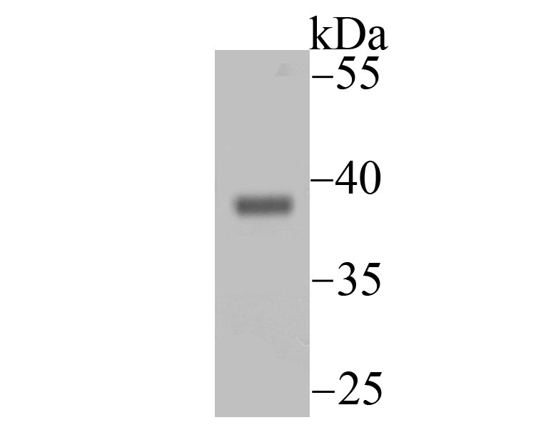
Fig1: Western blot analysis of Endo G on mouse heart tissue lysate using anti-Endo G antibody at 1/500 dilution.Application
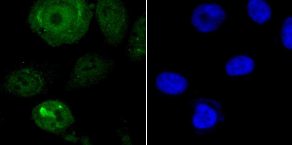
Fig2: ICC staining Endo G in PC-3M cells (green). The nuclear counter stain is DAPI (blue). Cells were fixed in paraformaldehyde, permeabilised with 0.25% Triton X100/PBS.Application
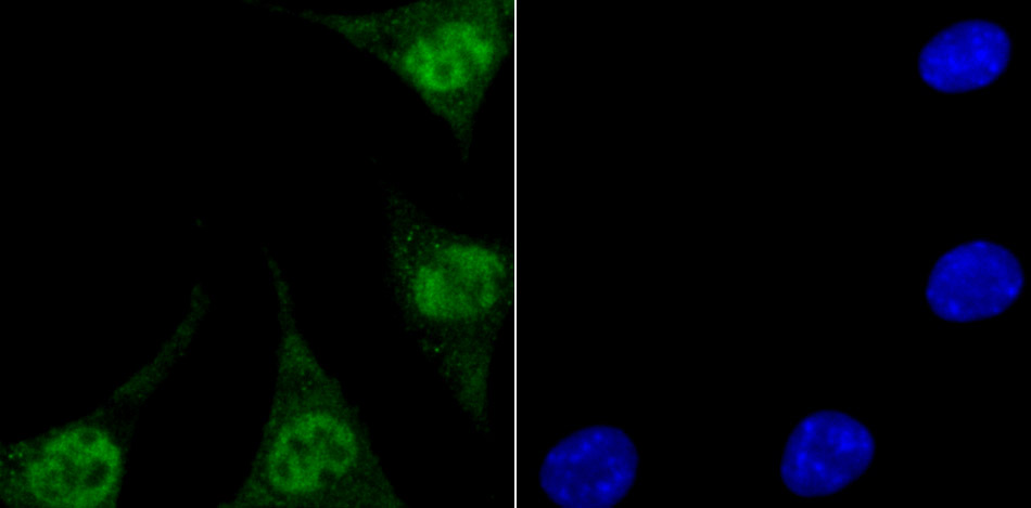
Fig3: ICC staining Endo G in SH-SY5Y cells (green). The nuclear counter stain is DAPI (blue). Cells were fixed in paraformaldehyde, permeabilised with 0.25% Triton X100/PBS.Application
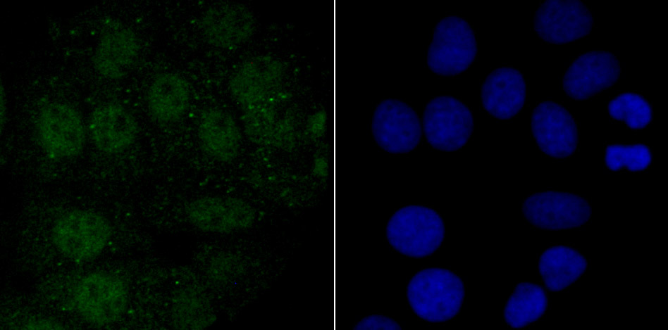
Fig4: ICC staining Endo G in MCF-7 cells (green). The nuclear counter stain is DAPI (blue). Cells were fixed in paraformaldehyde, permeabilised| Product Name | Endo G [JU31-30] |
|---|---|
| Antibody Type | Primary Antibodies |
| Antigen Alias | EndoG antibody Endonuclease G antibody Endonuclease G mitochondrial antibody EndonucleaseG antibody FLJ27463 antibody Mitochondrial endonuclease G antibody NUCG_HUMAN antibody |
| Product description | Endonuclease G (ENDOG), a nuclear encoded protein, localizes to the mitochondria. This sugar-nonspecific nuclease, responsible for major mitochondrial nuclease activity, preferentially cleaves single-stranded DNA(ssDNA). Synthesized as a propeptide with an amino-terminal presequence that targets the nuclease to mitochondria, ENDOG translocates to neuclei on apoptotic stimulation and act as a nuclease without sequence specificity. Both exonucleases and DNase I stimulate the ability of ENDOG to generate double-stranded DNA cleavage products at physiological ionic strengths, suggesting that these activities work in concert with ENDOG in apoptotic cells to ensure efficient DNA breakdown. In addition to deoxyribonuclease activities, ENDOG also has ribonuclease (RNase) and RNase H activities. ENDOG is capable of generating the RNA primers required by DNA polymerase gamma to initiate replication of mitochondrial DNA. ENDOG exists in the mitochondrial intermembrane space, but not in the matrix where mtDNA replication occurs. This enzyme provides an important nicking function for mitochondrial DNA specifically cleaving DNA at GC tracts. Human ENDOG maps to chromosome 9q34.11. |
| Immunogen | Recombinant protein |
| Clonality | Monoclonal |
|---|---|
| Isotype | IgG |
| Host Species | Recombinant rabbit |
| Tested Applications | WBICC |
| WB:1:500-1:1,000 ICC:1:50-1:200 |
|
| Species Reactivity | Human |
| Concentration | 1mg/ml |
| Alternative Names | EndoG antibody Endonuclease G antibody Endonuclease G mitochondrial antibody EndonucleaseG antibody FLJ27463 antibody Mitochondrial endonuclease G antibody NUCG_HUMAN antibody |
|---|---|
| Molecular Weight(MW) | 36 kDa |
| Cellular Localization | Mitochondrion. Nucleus. |
| SwissProt ID | Q14249 |
|---|
Application
Fig1: Western blot analysis of Endo G on mouse heart tissue lysate using anti-Endo G antibody at 1/500 dilution.
Application
Fig2: ICC staining Endo G in PC-3M cells (green). The nuclear counter stain is DAPI (blue). Cells were fixed in paraformaldehyde, permeabilised with 0.25% Triton X100/PBS.
Application
Fig3: ICC staining Endo G in SH-SY5Y cells (green). The nuclear counter stain is DAPI (blue). Cells were fixed in paraformaldehyde, permeabilised with 0.25% Triton X100/PBS.
Application
Fig4: ICC staining Endo G in MCF-7 cells (green). The nuclear counter stain is DAPI (blue). Cells were fixed in paraformaldehyde, permeabilised| Positive Control | MCF-7, PC-3M, SH-SY5Y, mouse heart tissue lysate. |
|---|---|
| Application Notes | WB:1:500-1:1,000 ICC:1:50-1:200 |
| Form | Liquid |
|---|---|
| Storage Instructions | Store at +4℃ after thawing. Aliquot store at -20℃ or -80℃. Avoid repeated freeze / thaw cycles. |
| Storage Buffer | 1*TBS (pH7.4), 1%BSA, 40%Glycerol. Preservative: 0.05% Sodium Azide. |


