Application
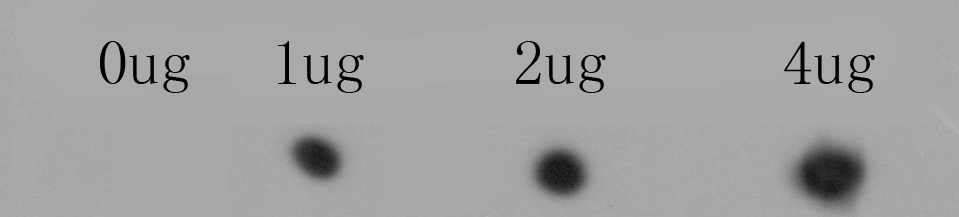
Fig1: Dot blot analysis of anti-RYR1 immunization peptide on PVDF. 1ug, 2ug and 4ug peptides were given in this test. Anti-RYR1 antibody was diluted with 1/500.Application
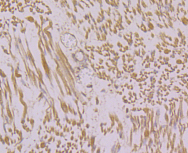
Fig2: Immunohistochemical analysis of paraffin-embedded human fetal skeletal muscle tissue using anti-RYR1 antibody. Counter stained with hematoxylin.Application
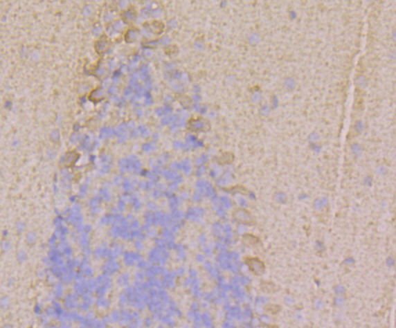
Fig3: Immunohistochemical analysis of paraffin-embedded mouse cerebellum tissue using anti-RYR1 antibody. Counter stained with hematoxylin.Application
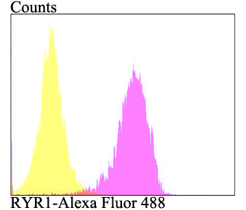
Fig4: Flow cytometric analysis of SiHa cells with RYR1 antibody at 1/100 dilution (fuchsia) compared with an unlabelled control (cells without incubation with primary antibody; yellow). Alexa Fluor 488-conjugated goat anti-rabbit IgG was used as the secondary antibody.| Product Name | RYR1 |
|---|---|
| Antibody Type | Primary Antibodies |
| Antigen Alias | Arrhythmogenic right ventricular dysplasia 2 antibody ARVC2 antibody ARVD2 antibody Brain ryanodine receptor calcium release channel antibody Brain type ryanodine receptor antibody Cardiac muscle ryanodine receptor calcium release channel antibody Cardiac muscle type ryanodine receptor antibody CCO antibody Central core disease of muscle antibody HBRR antibody hRYR 2 antibody hRYR2 antibody MHS antibody MHS1 antibody Ryanodine receptor 1 (skeletal) antibody Ryanodine receptor 1 antibody Ryanodine receptor 2 (cardiac) antibody Ryanodine receptor 2 antibody Ryanodine receptor 3 antibody Ryanodine receptor type1 antibody RYDR antibody RYR 1 antibody RYR 2 antibody RYR 3 antibody RYR antibody RYR-1 antibody RyR1 antibody RYR1_HUMAN antibody RYR2 antibody RYR3 antibody Sarcoplasmic reticulum calcium release channel antibody Skeletal muscle calcium release channel antibody Skeletal muscle ryanodine receptor antibody Skeletal muscle type ryanodine receptor antibody Skeletal muscle-type ryanodine receptor antibody SKRR antibody Type 1 like ryanodine receptor antibody Type 1 ryanodine receptor antibody VTSIP antibody |
| Product description | Calcium channel that mediates the release of Ca2+ from the sarcoplasmic reticulum into the cytoplasm and thereby plays a key role in triggering muscle contraction following depolarization of T-tubules. Repeated very high-level exercise increases the open probability of the channel and leads to Ca2+ leaking into the cytoplasm. Can also mediate the release of Ca2+ from intracellular stores in neurons, and may thereby promote prolonged Ca2+ signaling in the brain. Required for normal embryonic development of muscle fibers and skeletal muscle. Required for normal heart morphogenesis, skin development and ossification during embryogenesis. |
| Immunogen | Synthetic peptide within human RYR1 aa 1350-1400. |
| Clonality | Polyclonal |
|---|---|
| Isotype | IgG |
| Host Species | Rabbit |
| Tested Applications | DotIHCFC |
| Dot Blot:1:500-1:1,000 IHC:1:50-1:200 FC:1:50-1:100 |
|
| Species Reactivity | HumanMouse |
| Concentration | 1mg/ml |
| Alternative Names | Arrhythmogenic right ventricular dysplasia 2 antibody ARVC2 antibody ARVD2 antibody Brain ryanodine receptor calcium release channel antibody Brain type ryanodine receptor antibody Cardiac muscle ryanodine receptor calcium release channel antibody Cardiac muscle type ryanodine receptor antibody CCO antibody Central core disease of muscle antibody HBRR antibody hRYR 2 antibody hRYR2 antibody MHS antibody MHS1 antibody Ryanodine receptor 1 (skeletal) antibody Ryanodine receptor 1 antibody Ryanodine receptor 2 (cardiac) antibody Ryanodine receptor 2 antibody Ryanodine receptor 3 antibody Ryanodine receptor type1 antibody RYDR antibody RYR 1 antibody RYR 2 antibody RYR 3 antibody RYR antibody RYR-1 antibody RyR1 antibody RYR1_HUMAN antibody RYR2 antibody RYR3 antibody Sarcoplasmic reticulum calcium release channel antibody Skeletal muscle calcium release channel antibody Skeletal muscle ryanodine receptor antibody Skeletal muscle type ryanodine receptor antibody Skeletal muscle-type ryanodine receptor antibody SKRR antibody Type 1 like ryanodine receptor antibody Type 1 ryanodine receptor antibody VTSIP antibody |
|---|---|
| Molecular Weight(MW) | 565 kDa |
| Cellular Localization | Sarcoplasmic reticulum membrane. |
Application
Fig1: Dot blot analysis of anti-RYR1 immunization peptide on PVDF. 1ug, 2ug and 4ug peptides were given in this test. Anti-RYR1 antibody was diluted with 1/500.
Application
Fig2: Immunohistochemical analysis of paraffin-embedded human fetal skeletal muscle tissue using anti-RYR1 antibody. Counter stained with hematoxylin.
Application
Fig3: Immunohistochemical analysis of paraffin-embedded mouse cerebellum tissue using anti-RYR1 antibody. Counter stained with hematoxylin.
Application
Fig4: Flow cytometric analysis of SiHa cells with RYR1 antibody at 1/100 dilution (fuchsia) compared with an unlabelled control (cells without incubation with primary antibody; yellow). Alexa Fluor 488-conjugated goat anti-rabbit IgG was used as the secondary antibody.| Positive Control | Human fetal skeletal muscle tissue, mouse cerebellum tissue, SiHa. |
|---|---|
| Application Notes | Dot Blot:1:500-1:1,000 IHC:1:50-1:200 FC:1:50-1:100 |
| Form | Liquid |
|---|---|
| Storage Instructions | Store at +4℃ after thawing. Aliquot store at -20℃. Avoid repeated freeze / thaw cycles. |
| Storage Buffer | 1*TBS (pH7.4), 0.5%BSA, 50%Glycerol. Preservative: 0.05% Sodium Azide. |


