Application
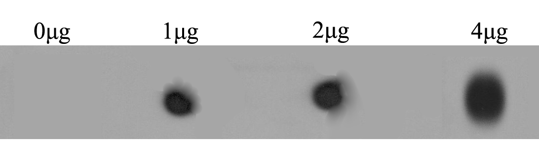
Fig1: Dot blot analysis of anti-RYR2 immunization peptide on PVDF. 1ug, 2ug and 4ug peptides were given in this test. Anti-RYR2 antibody was diluted with 1/500.Application
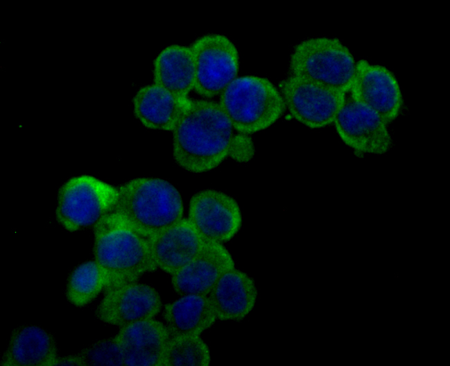
Fig2: ICC staining RYR2 in 293T cells (green). The nuclear counter stain is DAPI (blue). Cells were fixed in paraformaldehyde, permeabilised with 0.25% Triton X100/PBS.Application
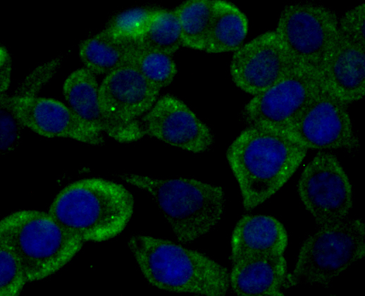
Fig3: ICC staining RYR2 in LOVO cells (green). The nuclear counter stain is DAPI (blue). Cells were fixed in paraformaldehyde, permeabilised with 0.25% Triton X100/PBS.Application
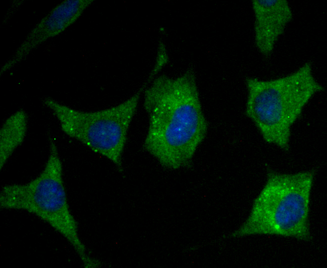
Fig4: ICC staining RYR2 in SH-SY-5Y cells (green). The nuclear counter stain is DAPI (blue). Cells were fixed in paraformaldehyde, permeabilised with 0.25% Triton X100/PBS.Application
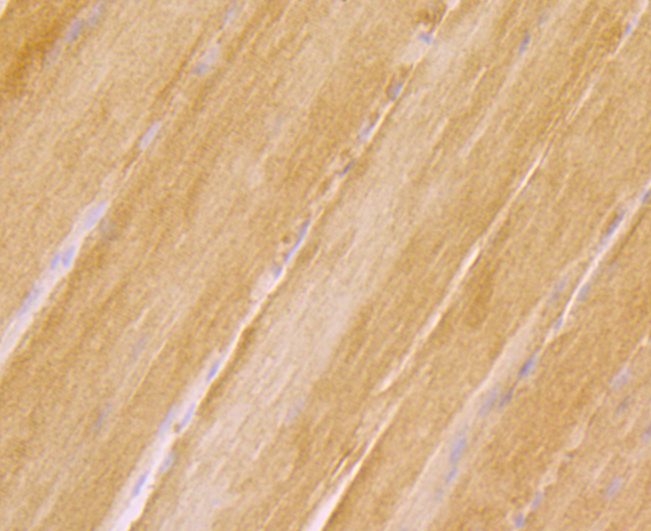
Fig5: Immunohistochemical analysis of paraffin-embedded rat skeletal muscle tissue using anti-RYR2 antibody. Counter stained with hematoxylin.Application
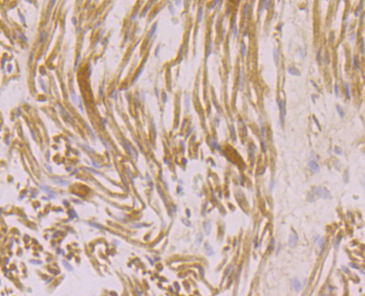
Fig6: Immunohistochemical analysis of paraffin-embedded human fetal skeletal muscle tissue using anti-RYR2 antibody. Counter stained with hematoxylin.Application
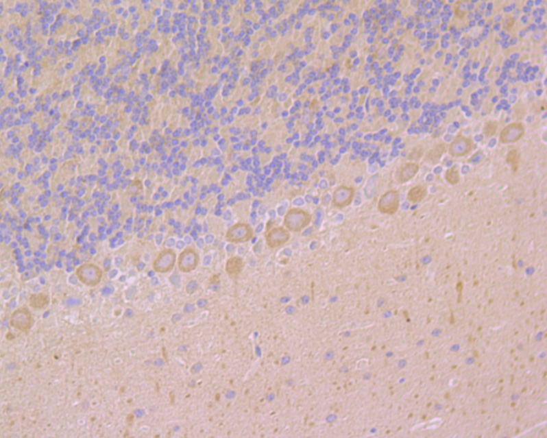
Fig7: Immunohistochemical analysis of paraffin-embedded mouse cerebellum tissue using anti-RYR2 antibody. Counter stained with hematoxylin.Application
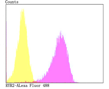
Fig8: Flow cytometric analysis of 293T cells with RYR2 antibody at 1/100 dilution (fuchsia) compared with an unlabelled control (cells without incubation with primary antibody; yellow). Alexa Fluor 488-conjugated goat anti-rabbit IgG was used as the secondary antibody.| Product Name | RYR2 |
|---|---|
| Antibody Type | Primary Antibodies |
| Antigen Alias | Arrhythmogenic right ventricular dysplasia 2 antibody ARVC2 antibody ARVD2 antibody Brain ryanodine receptor calcium release channel antibody Brain type ryanodine receptor antibody Cardiac muscle ryanodine receptor calcium release channel antibody Cardiac muscle type ryanodine receptor antibody CCO antibody Central core disease of muscle antibody HBRR antibody hRYR 2 antibody hRYR2 antibody MHS antibody MHS1 antibody Ryanodine receptor 1 (skeletal) antibody Ryanodine receptor 1 antibody Ryanodine receptor 2 (cardiac) antibody Ryanodine receptor 2 antibody Ryanodine receptor 3 antibody Ryanodine receptor type1 antibody RYDR antibody RYR 1 antibody RYR 2 antibody RYR 3 antibody RYR antibody RYR-1 antibody RyR1 antibody RYR1_HUMAN antibody RYR2 antibody RYR3 antibody Sarcoplasmic reticulum calcium release channel antibody Skeletal muscle calcium release channel antibody Skeletal muscle ryanodine receptor antibody Skeletal muscle type ryanodine receptor antibody Skeletal muscle-type ryanodine receptor antibody SKRR antibody Type 1 like ryanodine receptor antibody Type 1 ryanodine receptor antibody VTSIP antibody |
| Product description | Calcium channel that mediates the release of Ca2+ from the sarcoplasmic reticulum into the cytoplasm and thereby plays a key role in triggering cardiac muscle contraction. Aberrant channel activation can lead to cardiac arrhythmia. In cardiac myocytes, calcium release is triggered by increased Ca2+ levels due to activation of the L-type calcium channel CACNA1C. The calcium channel activity is modulated by formation of heterotetramers with RYR3. Required for cellular calcium ion homeostasis. Required for embryonic heart development. |
| Immunogen | Synthetic peptide within human RYR2 aa 1450-1550. |
| Clonality | Polyclonal |
|---|---|
| Isotype | IgG |
| Host Species | Rabbit |
| Tested Applications | WBICCIHCFC |
| WB:1:500 ICC:1:50-1:100 IHC:1:50-1:200 FC:1:50-1:100 |
|
| Species Reactivity | HumanMouseRat |
| Concentration | 1mg/ml |
| Alternative Names | Arrhythmogenic right ventricular dysplasia 2 antibody ARVC2 antibody ARVD2 antibody Brain ryanodine receptor calcium release channel antibody Brain type ryanodine receptor antibody Cardiac muscle ryanodine receptor calcium release channel antibody Cardiac muscle type ryanodine receptor antibody CCO antibody Central core disease of muscle antibody HBRR antibody hRYR 2 antibody hRYR2 antibody MHS antibody MHS1 antibody Ryanodine receptor 1 (skeletal) antibody Ryanodine receptor 1 antibody Ryanodine receptor 2 (cardiac) antibody Ryanodine receptor 2 antibody Ryanodine receptor 3 antibody Ryanodine receptor type1 antibody RYDR antibody RYR 1 antibody RYR 2 antibody RYR 3 antibody RYR antibody RYR-1 antibody RyR1 antibody RYR1_HUMAN antibody RYR2 antibody RYR3 antibody Sarcoplasmic reticulum calcium release channel antibody Skeletal muscle calcium release channel antibody Skeletal muscle ryanodine receptor antibody Skeletal muscle type ryanodine receptor antibody Skeletal muscle-type ryanodine receptor antibody SKRR antibody Type 1 like ryanodine receptor antibody Type 1 ryanodine receptor antibody VTSIP antibody |
|---|---|
| Molecular Weight(MW) | 565 kDa |
| Cellular Localization | Membrane. Sarcoplasmic reticulum. |
Application
Fig1: Dot blot analysis of anti-RYR2 immunization peptide on PVDF. 1ug, 2ug and 4ug peptides were given in this test. Anti-RYR2 antibody was diluted with 1/500.
Application
Fig2: ICC staining RYR2 in 293T cells (green). The nuclear counter stain is DAPI (blue). Cells were fixed in paraformaldehyde, permeabilised with 0.25% Triton X100/PBS.
Application
Fig3: ICC staining RYR2 in LOVO cells (green). The nuclear counter stain is DAPI (blue). Cells were fixed in paraformaldehyde, permeabilised with 0.25% Triton X100/PBS.
Application
Fig4: ICC staining RYR2 in SH-SY-5Y cells (green). The nuclear counter stain is DAPI (blue). Cells were fixed in paraformaldehyde, permeabilised with 0.25% Triton X100/PBS.
Application
Fig5: Immunohistochemical analysis of paraffin-embedded rat skeletal muscle tissue using anti-RYR2 antibody. Counter stained with hematoxylin.
Application
Fig6: Immunohistochemical analysis of paraffin-embedded human fetal skeletal muscle tissue using anti-RYR2 antibody. Counter stained with hematoxylin.
Application
Fig7: Immunohistochemical analysis of paraffin-embedded mouse cerebellum tissue using anti-RYR2 antibody. Counter stained with hematoxylin.
Application
Fig8: Flow cytometric analysis of 293T cells with RYR2 antibody at 1/100 dilution (fuchsia) compared with an unlabelled control (cells without incubation with primary antibody; yellow). Alexa Fluor 488-conjugated goat anti-rabbit IgG was used as the secondary antibody.| Positive Control | 293T, LOVO, SH-SY-5Y, human fetal skeletal muscle tissue, mouse brain tissue, mouse cerebellum tissue, rat skeletal muscle tissue. |
|---|---|
| Application Notes | WB:1:500 ICC:1:50-1:100 IHC:1:50-1:200 FC:1:50-1:100 |
| Form | Liquid |
|---|---|
| Storage Instructions | Store at +4℃ after thawing. Aliquot store at -20℃. Avoid repeated freeze / thaw cycles. |
| Storage Buffer | 1*TBS (pH7.4), 0.5%BSA, 50%Glycerol. Preservative: 0.05% Sodium Azide. |


