Application
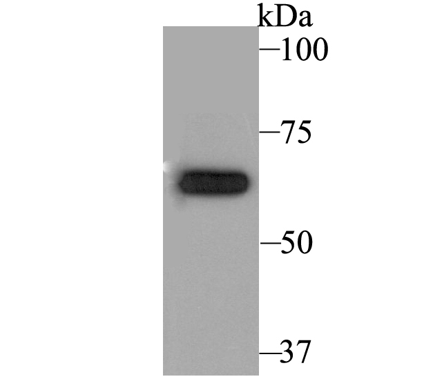
Fig1: Western blot analysis of KCNA5 on PANC-1 cell lysate using anti-KCNA5 antibody at 1/5,000 dilution.Application
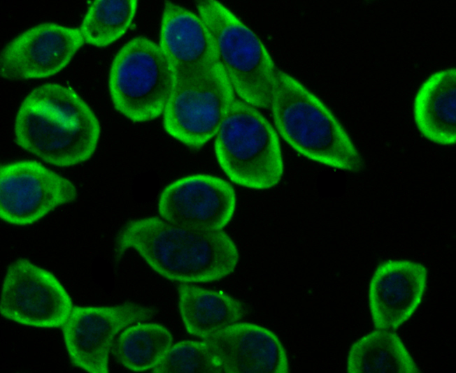
Fig2: ICC staining KCNA5 in LOVO cells (green). The nuclear counter stain is DAPI (blue). Cells were fixed in paraformaldehyde, permeabilised with 0.25% Triton X100/PBS.Application
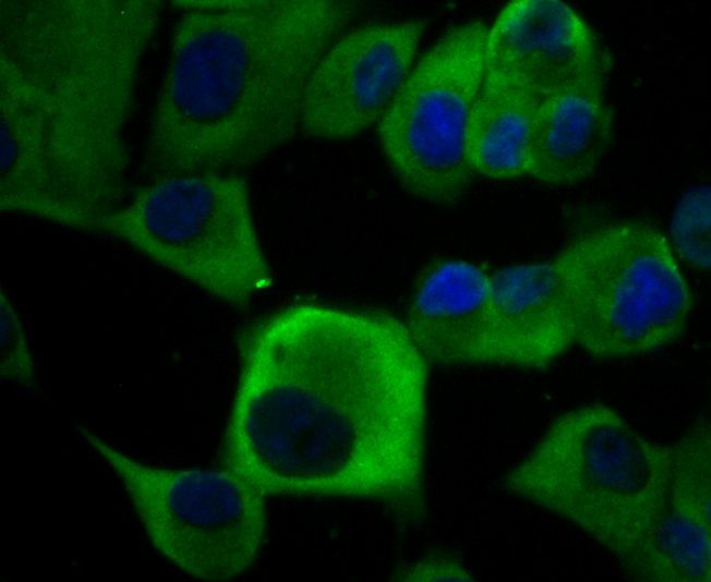
Fig3: ICC staining KCNA5 in PANC-1 cells (green). The nuclear counter stain is DAPI (blue). Cells were fixed in paraformaldehyde, permeabilised with 0.25% Triton X100/PBS.Application
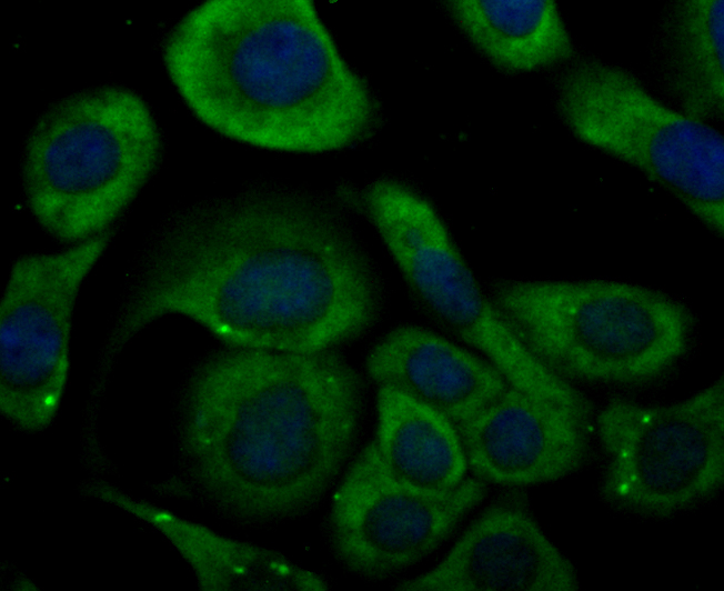
Fig4: ICC staining KCNA5 in SKOV-3 cells (green). The nuclear counter stain is DAPI (blue). Cells were fixed in paraformaldehyde, permeabilised with 0.25% Triton X100/PBS.Application
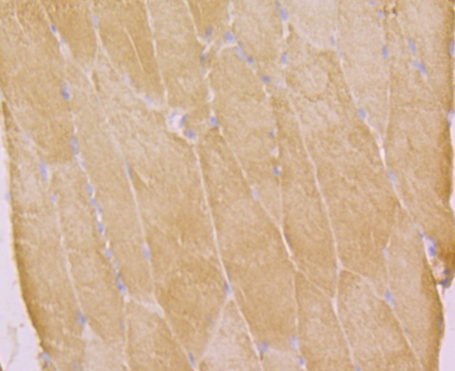
Fig5: Immunohistochemical analysis of paraffin-embedded rat skeletal muscle tissue using anti-KCNA5 antibody. Counter stained with hematoxylin.Application
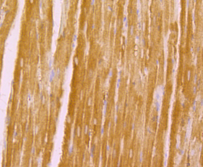
Fig6: Immunohistochemical analysis of paraffin-embedded rat heart tissue using anti-KCNA5 antibody. Counter stained with hematoxylin.Application
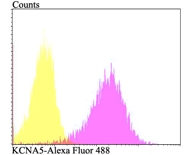
Fig7: Flow cytometric analysis of PANC-1 cells with KCNA5 antibody at 1/100 dilution (fuchsia) compared with an unlabelled control (cells without incubation with primary antibody; yellow). Alexa Fluor 488-conjugated goat anti-rabbit IgG was used as the secondary antibody.| Product Name | KCNA5 |
|---|---|
| Antibody Type | Primary Antibodies |
| Antigen Alias | ATFB7 antibody cardiac potassium channel antibody HCK1 antibody HK2 antibody HPCN1 antibody insulinoma and islet potassium channel antibody KCNA5 antibody KCNA5_HUMAN antibody KV1.5 antibody PCN1 antibody potassium channel 1 antibody Potassium channel, insulinoma and islet cell antibody Potassium channel, voltage-gated, shaker-related subfamily, member 5 antibody Potassium voltage gated channel subfamily A member 5 antibody Potassium voltage-gated channel subfamily A member 5 antibody Voltage-gated potassium channel HK2 antibody voltage-gated potassium channel protein Kv1.5 antibody Voltage-gated potassium channel subunit Kv1.5 antibody |
| Product description | Voltage-gated potassium channel that mediates transmembrane potassium transport in excitable membranes. Forms tetrameric potassium-selective channels through which potassium ions pass in accordance with their electrochemical gradient. The channel alternates between opened and closed conformations in response to the voltage difference across the membrane. Can form functional homotetrameric channels and heterotetrameric channels that contain variable proportions of KCNA1, KCNA2, KCNA4, KCNA5, and possibly other family members as well; channel properties depend on the type of alpha subunits that are part of the channel. Channel properties are modulated by cytoplasmic beta subunits that regulate the subcellular location of the alpha subunits and promote rapid inactivation. Homotetrameric channels display rapid activation and slow inactivation. May play a role in regulating the secretion of insulin in normal pancreatic islets. Isoform 2 exhibits a voltage-dependent recovery from inactivation and an excessive cumulative inactivation. |
| Immunogen | Synthetic peptide within rat KCNA5 aa 230-290. |
| Clonality | Polyclonal |
|---|---|
| Isotype | IgG |
| Host Species | Rabbit |
| Tested Applications | WBICCIHCFC |
| WB:1:1,000-1:5,000 ICC:1:50-1:200 IHC:1:50-1:200 FC:1:50-1:100 |
|
| Species Reactivity | HumanMouseRat |
| Concentration | 1mg/ml |
| Alternative Names | ATFB7 antibody
cardiac potassium channel antibody
HCK1 antibody
HK2 antibody
HPCN1 antibody
insulinoma and islet potassium channel antibody
KCNA5 antibody
KCNA5_HUMAN antibody
KV1.5 antibody
PCN1 antibody
potassium channel 1 antibody
Potassium channel insulinoma and islet cell antibody Potassium channel voltage-gated shaker-related subfamily member 5 antibody Potassium voltage gated channel subfamily A member 5 antibody Potassium voltage-gated channel subfamily A member 5 antibody Voltage-gated potassium channel HK2 antibody voltage-gated potassium channel protein Kv1.5 antibody Voltage-gated potassium channel subunit Kv1.5 antibody |
|---|---|
| Molecular Weight(MW) | 67 kDa |
| Cellular Localization | Plasma membrane. |
Application
Fig1: Western blot analysis of KCNA5 on PANC-1 cell lysate using anti-KCNA5 antibody at 1/5,000 dilution.
Application
Fig2: ICC staining KCNA5 in LOVO cells (green). The nuclear counter stain is DAPI (blue). Cells were fixed in paraformaldehyde, permeabilised with 0.25% Triton X100/PBS.
Application
Fig3: ICC staining KCNA5 in PANC-1 cells (green). The nuclear counter stain is DAPI (blue). Cells were fixed in paraformaldehyde, permeabilised with 0.25% Triton X100/PBS.
Application
Fig4: ICC staining KCNA5 in SKOV-3 cells (green). The nuclear counter stain is DAPI (blue). Cells were fixed in paraformaldehyde, permeabilised with 0.25% Triton X100/PBS.
Application
Fig5: Immunohistochemical analysis of paraffin-embedded rat skeletal muscle tissue using anti-KCNA5 antibody. Counter stained with hematoxylin.
Application
Fig6: Immunohistochemical analysis of paraffin-embedded rat heart tissue using anti-KCNA5 antibody. Counter stained with hematoxylin.
Application
Fig7: Flow cytometric analysis of PANC-1 cells with KCNA5 antibody at 1/100 dilution (fuchsia) compared with an unlabelled control (cells without incubation with primary antibody; yellow). Alexa Fluor 488-conjugated goat anti-rabbit IgG was used as the secondary antibody.| Positive Control | PANC-1, LOVO, SKOV-3, rat skeletal muscle tissue, rat heart tissue. |
|---|---|
| Application Notes | WB:1:1,000-1:5,000 ICC:1:50-1:200 IHC:1:50-1:200 FC:1:50-1:100 |
| Form | Liquid |
|---|---|
| Storage Instructions | Store at +4℃ after thawing. Aliquot store at -20℃. Avoid repeated freeze / thaw cycles. |
| Storage Buffer | 1*TBS (pH7.4), 0.5%BSA, 50%Glycerol. Preservative: 0.05% Sodium Azide. |


