Application
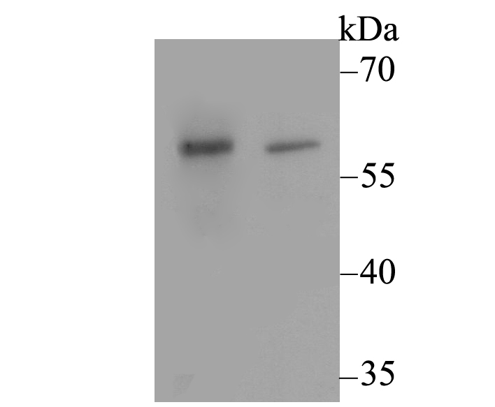
Fig1: Western blot analysis of CACNB3 on different lysates using anti-CACNB3 antibody at 1/500 dilution. Positive control: Lane1: Rat brain tissue Lane2: SKOV-3Application
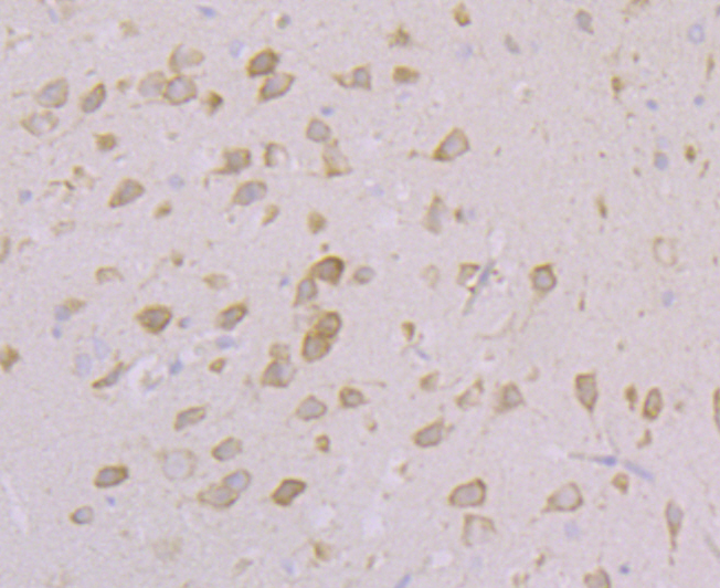
Fig2: Immunohistochemical analysis of paraffin-embedded rat brain tissue using anti-CACNB3 antibody. Counter stained with hematoxylin.Application
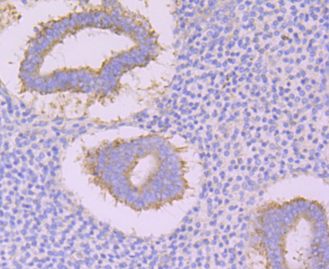
Fig3: Immunohistochemical analysis of paraffin-embedded human uterus tissue using anti-CACNB3 antibody. Counter stained with hematoxylin.Application
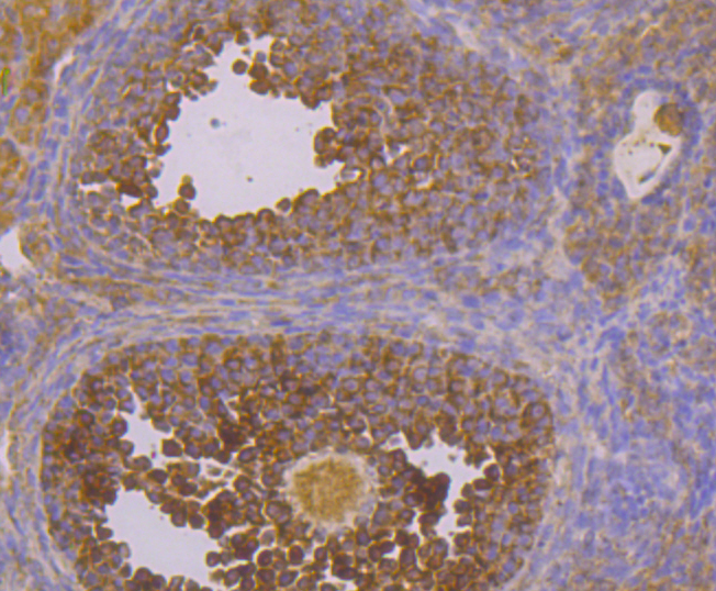
Fig4: Immunohistochemical analysis of paraffin-embedded mouse ovary tissue using anti-CACNB3 antibody. Counter stained with hematoxylin.Application
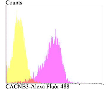
Fig5: Flow cytometric analysis of SKOV-3 cells with CACNB3 antibody at 1/100 dilution (fuchsia) compared with an unlabelled control (cells without incubation with primary antibody; yellow). Alexa Fluor 488-conjugated goat anti-rabbit IgG was used as the secondary antibody.| Product Name | CACNB3 |
|---|---|
| Antibody Type | Primary Antibodies |
| Antigen Alias | CAB3 antibody CACB3_HUMAN antibody Cacnb3 antibody CACNLB3 antibody Calcium channel voltage dependent beta 3 subunit antibody Calcium channel voltage-dependent subunit beta 3 antibody FLJ58949 antibody Voltage-dependent L-type calcium channel subunit beta-3 antibody |
| Product description | Regulatory subunit of the voltage-gated calcium channel that gives rise to L-type calcium currents. Increases CACNA1B peak calcium current and shifts the voltage dependencies of channel activation and inactivation. Increases CACNA1C peak calcium current and shifts the voltage dependencies of channel activation and inactivation. |
| Immunogen | Synthetic peptide within C-terminal human CACNB3. |
| Clonality | Polyclonal |
|---|---|
| Isotype | IgG |
| Host Species | Rabbit |
| Tested Applications | WBIHCFC |
| WB:1:500 IHC:1:50-1:200 FC:1:50-1:100 |
|
| Species Reactivity | HumanMouseRat |
| Concentration | 1mg/ml |
| Alternative Names | CAB3 antibody CACB3_HUMAN antibody Cacnb3 antibody CACNLB3 antibody Calcium channel voltage dependent beta 3 subunit antibody Calcium channel voltage-dependent subunit beta 3 antibody FLJ58949 antibody Voltage-dependent L-type calcium channel subunit beta-3 antibody |
|---|---|
| Molecular Weight(MW) | 55 kDa (Predicted band size) |
| Cellular Localization | Cytoplasm. |
Application
Fig1: Western blot analysis of CACNB3 on different lysates using anti-CACNB3 antibody at 1/500 dilution. Positive control: Lane1: Rat brain tissue Lane2: SKOV-3
Application
Fig2: Immunohistochemical analysis of paraffin-embedded rat brain tissue using anti-CACNB3 antibody. Counter stained with hematoxylin.
Application
Fig3: Immunohistochemical analysis of paraffin-embedded human uterus tissue using anti-CACNB3 antibody. Counter stained with hematoxylin.
Application
Fig4: Immunohistochemical analysis of paraffin-embedded mouse ovary tissue using anti-CACNB3 antibody. Counter stained with hematoxylin.
Application
Fig5: Flow cytometric analysis of SKOV-3 cells with CACNB3 antibody at 1/100 dilution (fuchsia) compared with an unlabelled control (cells without incubation with primary antibody; yellow). Alexa Fluor 488-conjugated goat anti-rabbit IgG was used as the secondary antibody.| Positive Control | Rat brain tissue lysate, SKOV-3, human uterus tissue, mouse ovary tissue. |
|---|---|
| Application Notes | WB:1:500 IHC:1:50-1:200 FC:1:50-1:100 |
| Form | Liquid |
|---|---|
| Storage Instructions | Store at +4℃ after thawing. Aliquot store at -20℃. Avoid repeated freeze / thaw cycles. |
| Storage Buffer | 1*TBS (pH7.4), 0.5%BSA, 50%Glycerol. Preservative: 0.05% Sodium Azide. |


