Application
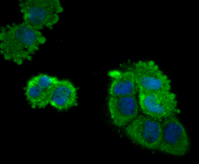
Fig1: ICC staining Eg5 in A431 cells (green). The nuclear counter stain is DAPI (blue). Cells were fixed in paraformaldehyde, permeabilised with 0.25% Triton X100/PBS.Application
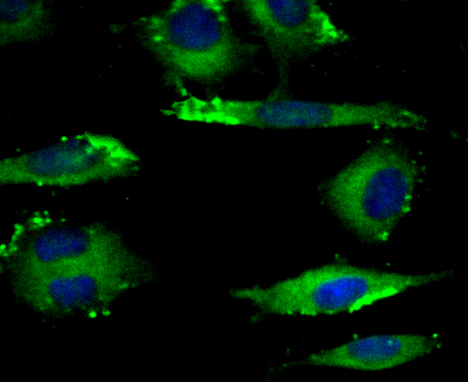
Fig2: ICC staining Eg5 in SH-SY-5Y cells (green). The nuclear counter stain is DAPI (blue). Cells were fixed in paraformaldehyde, permeabilised with 0.25% Triton X100/PBS.Application
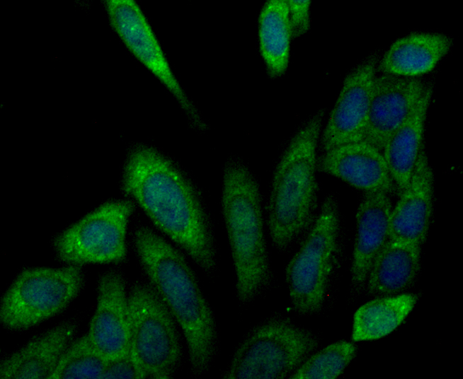
Fig3: ICC staining Eg5 in SiHa cells (green). The nuclear counter stain is DAPI (blue). Cells were fixed in paraformaldehyde, permeabilised with 0.25% Triton X100/PBS.Application
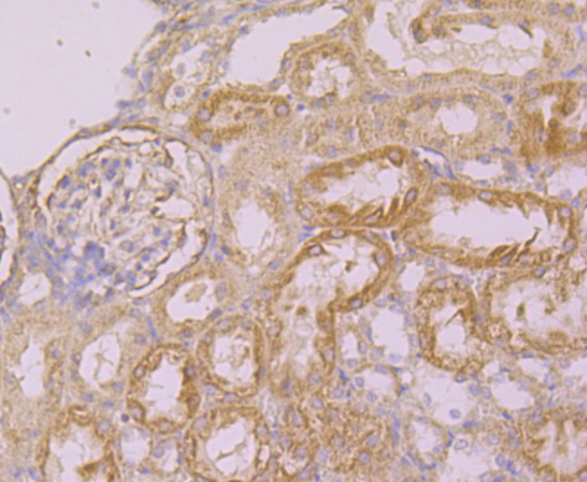
Fig4: Immunohistochemical analysis of paraffin-embedded rat kidney tissue using anti-Eg5 antibody. Counter stained with hematoxylin.Application
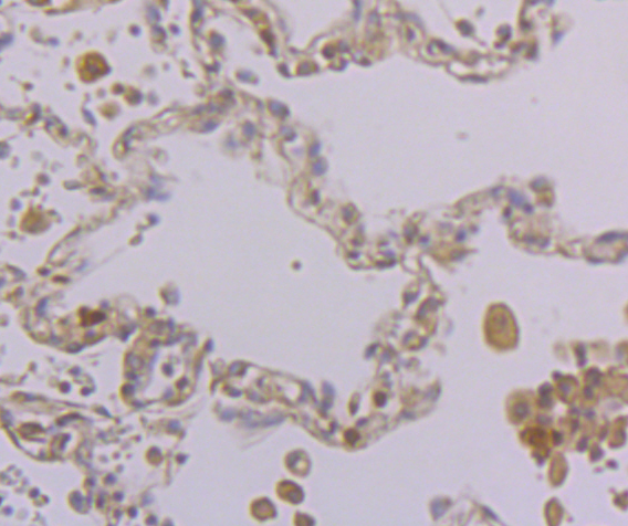
Fig5: Immunohistochemical analysis of paraffin-embedded human lung cancer tissue using anti-Eg5 antibody. Counter stained with hematoxylin.Application
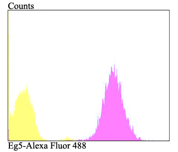
Fig6: Flow cytometric analysis of A431 cells with Eg5 antibody at 1/100 dilution (fuchsia) compared with an unlabelled control (cells without incubation with primary antibody; yellow). Alexa Fluor 488-conjugated goat anti-rabbit IgG was used as the secondary antibody.| Product Name | Eg5 |
|---|---|
| Antibody Type | Primary Antibodies |
| Antigen Alias | EG5 antibody HKSP antibody KIF11 antibody KIF11_HUMAN antibody Kinesin family member 11 antibody Kinesin like protein 1 antibody Kinesin-like protein 1 antibody Kinesin-like protein KIF11 antibody Kinesin-like spindle protein HKSP antibody Kinesin-related motor protein Eg5 antibody KNSL1 antibody MCLMR antibody Thyroid receptor-interacting protein 5 antibody TR-interacting protein 5 antibody TRIP-5 antibody TRIP5 antibody |
| Product description | Eukaryotes contain a superfamily of microtubule-based motor proteins comprising kinesin and a number of related proteins that are thought to participate in various forms of intracellular motility, including cell division and organelle transport. KIF11(also known as kinesin family member 11, Eg5 or TRIP5) is a slow, plus-end-directed microtubule-based motor of the BimC kinesin family that is essential for bipolar spindle formation during eukaryotic cell division. When the expression of KIF11 is blocked, centrosome migration halts and cells are arrested in mitosis with monoastral microtubule arrays. KIF11 is phosphorylated on serine during S phase and on both serine and Thr 927 during mitosis, which regulates the association of Eg5 with the spindle apparatus (probably during early prophase). KIF11 is also known to be a member of the thyroid receptor interacting protein (Trip) family, and interacts with the thyroid hormone receptor only in the presence of thyroid hormone. |
| Immunogen | Recombinant protein within human Eg5 aa 100-300. |
| Clonality | Polyclonal |
|---|---|
| Isotype | IgG |
| Host Species | Rabbit |
| Tested Applications | ICCIHCFC |
| ICC:1:50 IHC:1:50-1:200 FC:1:50-1:100 |
|
| Species Reactivity | HumanMouseRat |
| Concentration | 1mg/ml |
| Alternative Names | EG5 antibody HKSP antibody KIF11 antibody KIF11_HUMAN antibody Kinesin family member 11 antibody Kinesin like protein 1 antibody Kinesin-like protein 1 antibody Kinesin-like protein KIF11 antibody Kinesin-like spindle protein HKSP antibody Kinesin-related motor protein Eg5 antibody KNSL1 antibody MCLMR antibody Thyroid receptor-interacting protein 5 antibody TR-interacting protein 5 antibody TRIP-5 antibody TRIP5 antibody |
|---|---|
| Molecular Weight(MW) | 119 kDa |
| Cellular Localization | Cytoplasm, Cytoskeleton. |
Application
Fig1: ICC staining Eg5 in A431 cells (green). The nuclear counter stain is DAPI (blue). Cells were fixed in paraformaldehyde, permeabilised with 0.25% Triton X100/PBS.
Application
Fig2: ICC staining Eg5 in SH-SY-5Y cells (green). The nuclear counter stain is DAPI (blue). Cells were fixed in paraformaldehyde, permeabilised with 0.25% Triton X100/PBS.
Application
Fig3: ICC staining Eg5 in SiHa cells (green). The nuclear counter stain is DAPI (blue). Cells were fixed in paraformaldehyde, permeabilised with 0.25% Triton X100/PBS.
Application
Fig4: Immunohistochemical analysis of paraffin-embedded rat kidney tissue using anti-Eg5 antibody. Counter stained with hematoxylin.
Application
Fig5: Immunohistochemical analysis of paraffin-embedded human lung cancer tissue using anti-Eg5 antibody. Counter stained with hematoxylin.
Application
Fig6: Flow cytometric analysis of A431 cells with Eg5 antibody at 1/100 dilution (fuchsia) compared with an unlabelled control (cells without incubation with primary antibody; yellow). Alexa Fluor 488-conjugated goat anti-rabbit IgG was used as the secondary antibody.| Positive Control | A431, SH-SY-5Y, SiHa, rat kidney tissue, human lung cancer tissue. |
|---|---|
| Application Notes | ICC:1:50 IHC:1:50-1:200 FC:1:50-1:100 |
| Form | Liquid |
|---|---|
| Storage Instructions | Store at +4℃ after thawing. Aliquot store at -20℃. Avoid repeated freeze / thaw cycles. |
| Storage Buffer | 1*TBS (pH7.4), 0.5%BSA, 50%Glycerol. Preservative: 0.05% Sodium Azide. |


