Application
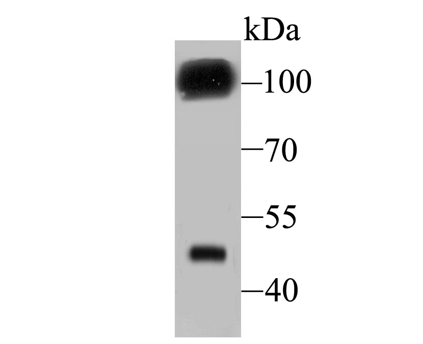
Fig1: Western blot analysis of GABA B Receptor 2 on mouse cerebellum tissue lysate using anti- GABA B Receptor 2 antibody at 1/500 dilution.Application
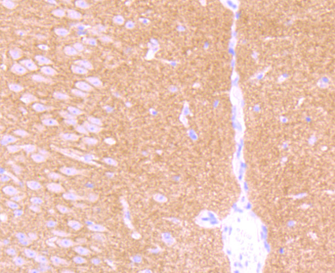
Fig2: Immunohistochemical analysis of paraffin-embedded mouse brain tissue using anti- GABA B Receptor 2 antibody. Counter stained with hematoxylin.Application
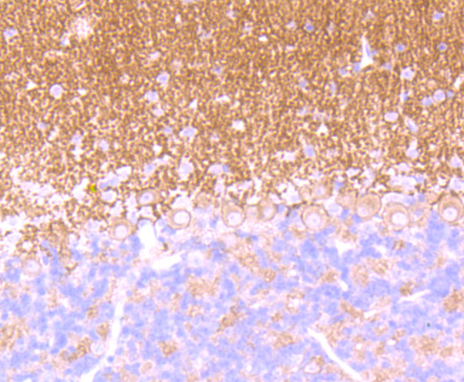
Fig3: Immunohistochemical analysis of paraffin-embedded mouse cerebellum tissue using anti- GABA B Receptor 2 antibody. Counter stained with hematoxylin.Application
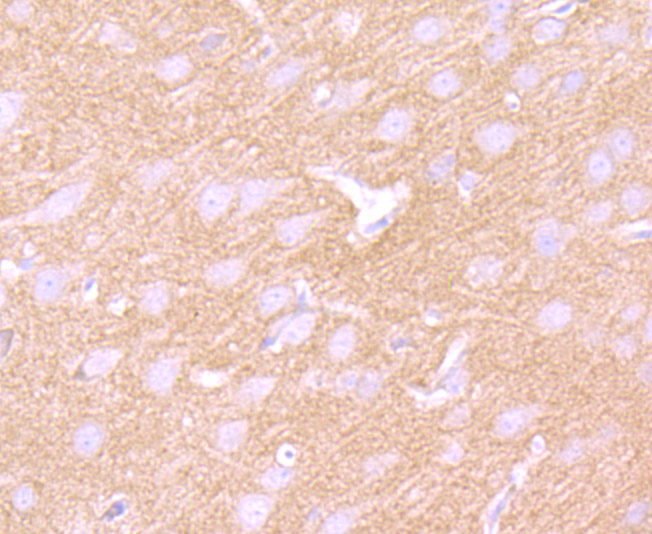
Fig4: Immunohistochemical analysis of paraffin-embedded rat brain tissue using anti- GABA B Receptor 2 antibody. Counter stained with hematoxylin.Application
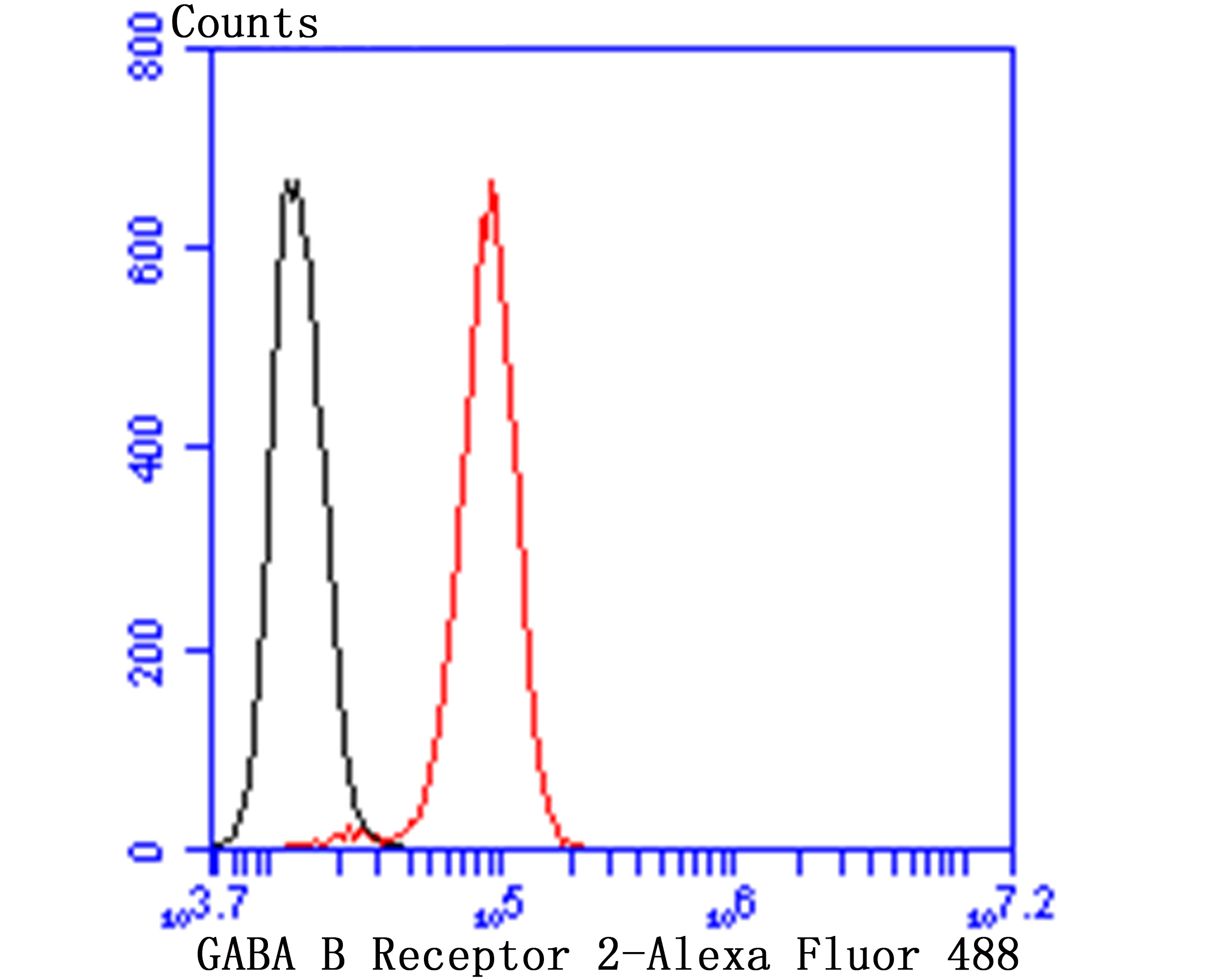
Fig5: Flow cytometric analysis of SH-SY-5Y cells with GABA B Receptor 2 antibody at 1/50 dilution (red) compared with an unlabelled control (cells without incubation with primary antibody; black). Alexa Fluor 488-conjugated goat anti rabbit IgG was used as the secondary antibody.| Product Name | GABA B Receptor 2 [JU31-32] |
|---|---|
| Antibody Type | Primary Antibodies |
| Antigen Alias | BcDNA:GH07312 antibody CG6706 antibody CT20836 antibody D Gaba2 antibody FLJ36928 antibody G protein coupled receptor 51 antibody G-protein coupled receptor 51 antibody GAB B R2 antibody GABA B R2 antibody GABA B receptor 2 antibody GABA-B receptor 2 antibody GABA-B-R2 antibody GABA-BR2 antibody GABAB R2 antibody GABABR 2 antibody GABABR2 antibody GABB R2 antibody GABBR 2 antibody Gabbr2 antibody GABR2_HUMAN antibody Gamma aminobutyric acid B receptor 2 antibody Gamma aminobutyric acid GABA B receptor 2 antibody Gamma aminobutyric acid type B receptor subunit 2 antibody Gamma-aminobutyric acid type B receptor subunit 2 antibody Gb 2 antibody Gb2 antibody GH07312 antibody GPR 51 antibody GPR51 antibody GPRC 3B antibody GPRC3B antibody HG 20 antibody HG20 antibody HRIHFB2099 antibody Metabotropic GABA B receptor subtype 2 antibody OTTHUMP00000021776 antibody OTTHUMP00000063797 antibody R2 SUBUNIT antibody |
| Product description | In the central nervous system (CNS), gamma-aminobutyric acid (GABA) is the main main inhibitory neurotransmitter that functions to regulate neuronal firing. GABA exerts its effects through two different kinds of receptors: ionotropic receptors (GABAA R and GABAC R), which produce fast inhibitory signals, and metabotropic receptors (GABAB R), which produce slow inhibitory signals. The GABAB R receptor is a heterodimer that consists of two multi-pass membrane proteins, designated GABAB R1 and GABAB R2, both of which belong to the G protein-coupled receptor family and are highly expressed in brain tissue. Together, GABAB R1 and GABAB R2 play a crucial role in the fine-tuning of inhibitory synaptic transmissions and are implicated in slow wave sleep, muscle relaxation, hippocampal long-term potentiation and antinociception events. Both GABAB R1 and GABAB R2 are regulated by G proteins that have a variety of functions, including activation of potassium channels, inhibition of adenylyl cyclase (A cyclase) activity and modulation of inositol phospholipid hydrolysis. |
| Immunogen | Recombinant protein |
| Clonality | Monoclonal |
|---|---|
| Isotype | IgG |
| Host Species | Recombinant rabbit |
| Tested Applications | WBIHCFCICC |
| WB:1:500-1:1,000 IHC:1:50-1:200 FC:1:50-1:100 ICC:1:50-1:100 |
|
| Species Reactivity | HumanMouseRat |
| Concentration | 1mg/ml |
| Alternative Names | BcDNA:GH07312 antibody CG6706 antibody CT20836 antibody D Gaba2 antibody FLJ36928 antibody G protein coupled receptor 51 antibody G-protein coupled receptor 51 antibody GAB B R2 antibody GABA B R2 antibody GABA B receptor 2 antibody GABA-B receptor 2 antibody GABA-B-R2 antibody GABA-BR2 antibody GABAB R2 antibody GABABR 2 antibody GABABR2 antibody GABB R2 antibody GABBR 2 antibody Gabbr2 antibody GABR2_HUMAN antibody Gamma aminobutyric acid B receptor 2 antibody Gamma aminobutyric acid GABA B receptor 2 antibody Gamma aminobutyric acid type B receptor subunit 2 antibody Gamma-aminobutyric acid type B receptor subunit 2 antibody Gb 2 antibody Gb2 antibody GH07312 antibody GPR 51 antibody GPR51 antibody GPRC 3B antibody GPRC3B antibody HG 20 antibody HG20 antibody HRIHFB2099 antibody Metabotropic GABA B receptor subtype 2 antibody OTTHUMP00000021776 antibody OTTHUMP00000063797 antibody R2 SUBUNIT antibody |
|---|---|
| Molecular Weight(MW) | 106 kDa |
| Cellular Localization | Plasma membrane. |
Application
Fig1: Western blot analysis of GABA B Receptor 2 on mouse cerebellum tissue lysate using anti- GABA B Receptor 2 antibody at 1/500 dilution.
Application
Fig2: Immunohistochemical analysis of paraffin-embedded mouse brain tissue using anti- GABA B Receptor 2 antibody. Counter stained with hematoxylin.
Application
Fig3: Immunohistochemical analysis of paraffin-embedded mouse cerebellum tissue using anti- GABA B Receptor 2 antibody. Counter stained with hematoxylin.
Application
Fig4: Immunohistochemical analysis of paraffin-embedded rat brain tissue using anti- GABA B Receptor 2 antibody. Counter stained with hematoxylin.
Application
Fig5: Flow cytometric analysis of SH-SY-5Y cells with GABA B Receptor 2 antibody at 1/50 dilution (red) compared with an unlabelled control (cells without incubation with primary antibody; black). Alexa Fluor 488-conjugated goat anti rabbit IgG was used as the secondary antibody.| Positive Control | Rat brain tissue, mouse brain tissue, mouse cerebellum tissue, SH-SY-5Y. |
|---|---|
| Application Notes | WB:1:500-1:1,000 IHC:1:50-1:200 FC:1:50-1:100 ICC:1:50-1:100 |
| Form | Liquid |
|---|---|
| Storage Instructions | Store at +4℃ after thawing. Aliquot store at -20℃ or -80℃. Avoid repeated freeze / thaw cycles. |
| Storage Buffer | 1*TBS (pH7.4), 1%BSA, 40%Glycerol. Preservative: 0.05% Sodium Azide. |


