Application
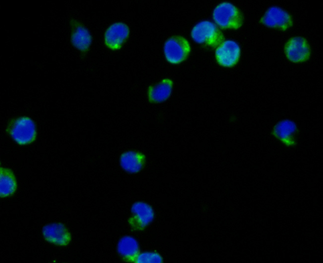
Fig1: ICC staining CD42b in Jurkat cells (green). The nuclear counter stain is DAPI (blue). Cells were fixed in paraformaldehyde, permeabilised with 0.25% Triton X100/PBS.Application
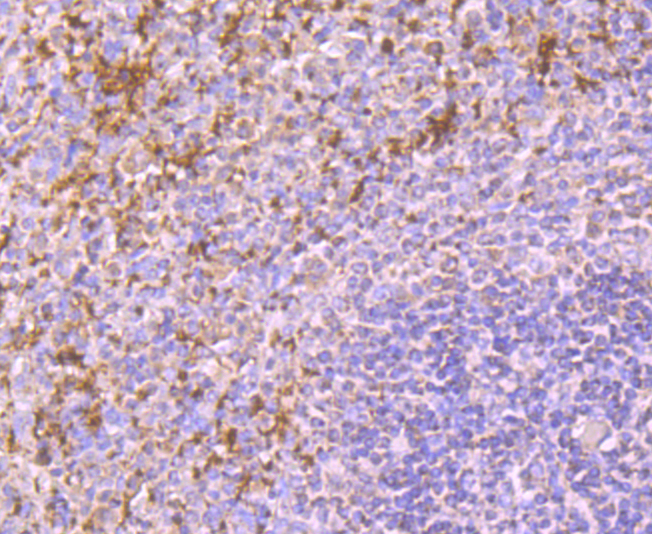
Fig2: Immunohistochemical analysis of paraffin-embedded human spleen tissue using anti-CD42b antibody. Counter stained with hematoxylin.Application
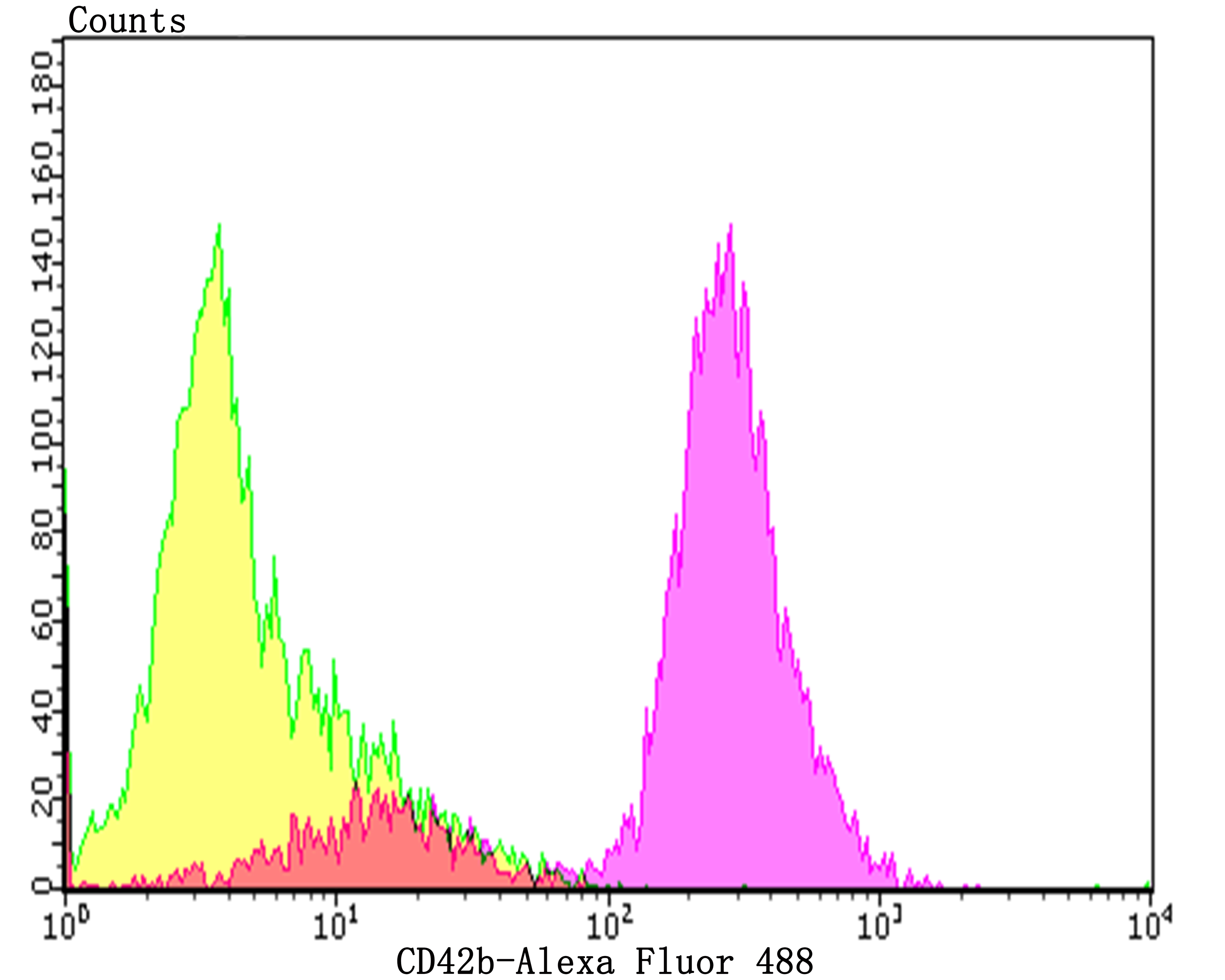
Fig3: Flow cytometric analysis of Jurkat cells with CD42b antibody at 1/50 dilution (purple) compared with an unlabelled control (cells without incubation with primary antibody; yellow). Alexa Fluor 488-conjugated goat anti-rabbit IgG was used as the secondary antibody.| Product Name | CD42b |
|---|---|
| Antibody Type | Primary Antibodies |
| Antigen Alias | Antigen CD42b alpha antibody BDPLT1 antibody BDPLT3 antibody BSS antibody CD 42b antibody CD42b alpha antibody CD42B antibody CD42b antigen antibody DBPLT3 antibody GLYCOCALICIN antibody Glycoprotein Ib (platelet) alpha polypeptide antibody Glycoprotein Ibalpha antibody GP Ib alpha antibody GP1B antibody GP1BA antibody GPIb alpha antibody GPIbA antibody MGC34595 antibody Platelet glycoprotein Ib alpha chain antibody Platelet glycoprotein Ib alpha polypeptide antibody Platelet membrane glycoprotein 1b alpha subunit antibody VWDP antibody |
| Product description | CD42a is a single-chain membrane glycoprotein that forms a noncovalent complex with CD42b. CD42b, also known as glycoprotein Ib a (GPIb a) is a membrane glycoprotein that is composed of a and b chains. The CD42b b chain is also designated CD42c, and is expressed on platelets and megakaryocytes. CD42a and CD42b are also present on platelets and mega-karyocytes, and the complex is a major component of the platelet surface. The complex acts as a receptor for von Willebrand's factor and as a von Willebrand's factor-dependent adhesion receptor. |
| Immunogen | Recombinant protein with human CD42b aa 130-310. |
| Clonality | Polyclonal |
|---|---|
| Isotype | IgG |
| Host Species | Rabbit |
| Tested Applications | WBICCIHCFC |
| ICC:1:50-1:200 IHC:1:50-1:200 FC:1:50-1:100 WB:1:500 |
|
| Species Reactivity | Human |
| Concentration | 1mg/ml |
| Alternative Names | Antigen CD42b alpha antibody BDPLT1 antibody BDPLT3 antibody BSS antibody CD 42b antibody CD42b alpha antibody CD42B antibody CD42b antigen antibody DBPLT3 antibody GLYCOCALICIN antibody Glycoprotein Ib (platelet) alpha polypeptide antibody Glycoprotein Ibalpha antibody GP Ib alpha antibody GP1B antibody GP1BA antibody GPIb alpha antibody GPIbA antibody MGC34595 antibody Platelet glycoprotein Ib alpha chain antibody Platelet glycoprotein Ib alpha polypeptide antibody Platelet membrane glycoprotein 1b alpha subunit antibody VWDP antibody |
|---|---|
| Molecular Weight(MW) | 71 kDa (Predicted band size) |
| Cellular Localization | Membrane. Secreted. |
| SwissProt ID | P07359 |
|---|
Application
Fig1: ICC staining CD42b in Jurkat cells (green). The nuclear counter stain is DAPI (blue). Cells were fixed in paraformaldehyde, permeabilised with 0.25% Triton X100/PBS.
Application
Fig2: Immunohistochemical analysis of paraffin-embedded human spleen tissue using anti-CD42b antibody. Counter stained with hematoxylin.
Application
Fig3: Flow cytometric analysis of Jurkat cells with CD42b antibody at 1/50 dilution (purple) compared with an unlabelled control (cells without incubation with primary antibody; yellow). Alexa Fluor 488-conjugated goat anti-rabbit IgG was used as the secondary antibody.| Positive Control | Jurkat, human spleen tissue. |
|---|---|
| Application Notes | ICC:1:50-1:200 IHC:1:50-1:200 FC:1:50-1:100 WB:1:500 |
| Form | Liquid |
|---|---|
| Storage Instructions | Store at +4℃ after thawing. Aliquot store at -20℃. Avoid repeated freeze / thaw cycles. |
| Storage Buffer | 1*TBS (pH7.4), 0.5%BSA, 50%Glycerol. Preservative: 0.05% Sodium Azide. |


