Application
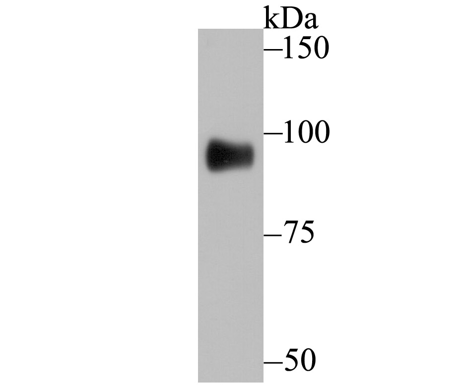
Fig1: Western blot analysis of GIT1 on A549 cell using anti-GIT1 antibody at 1/500 dilution.Application
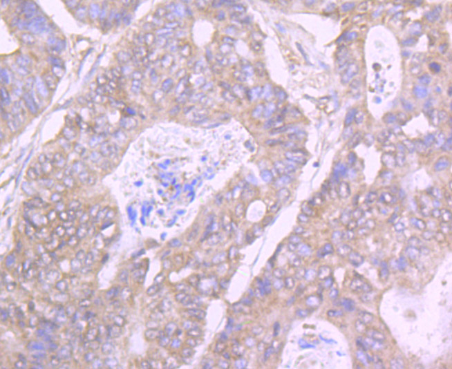
Fig2: Immunohistochemical analysis of paraffin-embedded human colon cancer tissue using anti-GIT1 antibody. Counter stained with hematoxylin.Application
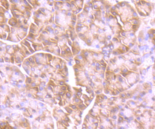
Fig3: Immunohistochemical analysis of paraffin-embedded human pancreas tissue using anti-GIT1 antibody. Counter stained with hematoxylin.Application
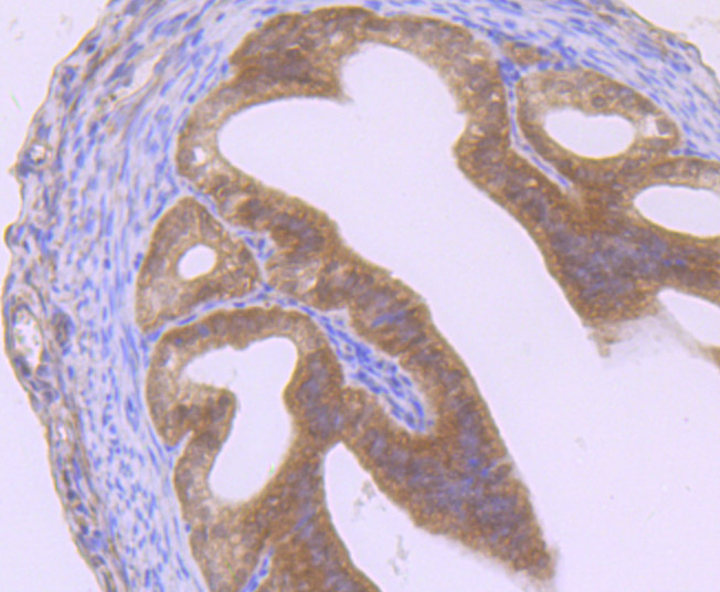
Fig4: Immunohistochemical analysis of paraffin-embedded mouse fallopian tubes tissue using anti-GIT1 antibody. Counter stained with hematoxylin.Application
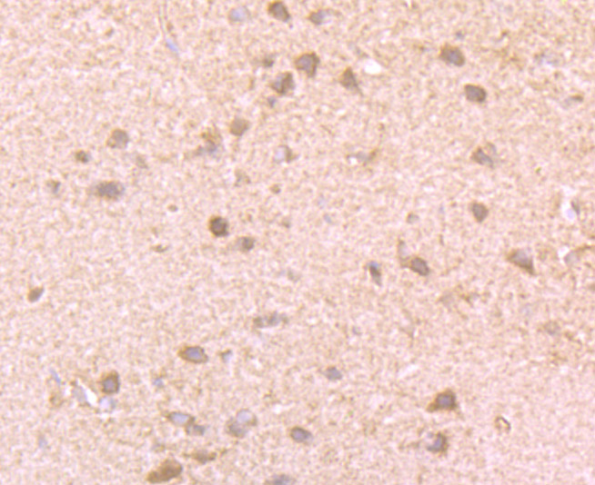
Fig5: Immunohistochemical analysis of paraffin-embedded rat brain tissue using anti-GIT1 antibody. Counter stained with hematoxylin.Application
Fig6: Flow cytometric analysis of A549 cells with GIT1 antibody at 1/100 dilution (red) compared with an unlabelled control (cells without incubation with primary antibody; black).| Product Name | GIT1 [JU33-39] |
|---|---|
| Antibody Type | Primary Antibodies |
| Antigen Alias | ARF GAP GIT1 antibody ARF GTPase activating protein GIT1 antibody ARF GTPase-activating protein GIT1 antibody CAT 1 antibody CAT-1 antibody CaT1 antibody Cool associated and tyrosine phosphorylated protein 1 antibody Cool-associated and tyrosine-phosphorylated protein 1 antibody G protein coupled receptor kinase interacting ArfGAP 1 antibody G protein coupled receptor kinase interactor 1 antibody G protein-coupled receptor kinase-interactor 1 antibody GIT1 antibody GIT1_HUMAN antibody GRK interacting protein 1 antibody GRK-interacting protein 1 antibody |
| Product description | Heterotrimeric G protein-mediated signal transduction is a dynamically regulated process with the intensity of signal decreasing over time despite the continued presence of the agonist. G protein-coupled receptor kinases (GRKs) are activated by activated G protein-coupled receptors, and they function to phosphorylate and inactivate cell surface receptors in the heterotrimeric G protein signaling cascade. GIT1 (for GRK-interactor 1) and GIT2 are GTPase-activating proteins (GAP) for members of the ADP ribosylation factor (ARF) family of small GTP-binding proteins, which are involved in vesicular trafficking. GIT1 overexpression results in reduced internalization and resensitization of b2-adrenergic receptor, thus reducing b2-adrenergic receptor signaling. |
| Immunogen | Recombinant protein |
| Clonality | Monoclonal |
|---|---|
| Isotype | IgG |
| Host Species | Recombinant rabbit |
| Tested Applications | WBIHCFC |
| WB:1:500-1:1,000 IHC:1:50-1:200 FC:1:50-1:100 |
|
| Species Reactivity | HumanMouseRat |
| Concentration | 1mg/ml |
| Alternative Names | ARF GAP GIT1 antibody ARF GTPase activating protein GIT1 antibody ARF GTPase-activating protein GIT1 antibody CAT 1 antibody CAT-1 antibody CaT1 antibody Cool associated and tyrosine phosphorylated protein 1 antibody Cool-associated and tyrosine-phosphorylated protein 1 antibody G protein coupled receptor kinase interacting ArfGAP 1 antibody G protein coupled receptor kinase interactor 1 antibody G protein-coupled receptor kinase-interactor 1 antibody GIT1 antibody GIT1_HUMAN antibody GRK interacting protein 1 antibody GRK-interacting protein 1 antibody |
|---|---|
| Molecular Weight(MW) | 85 kDa |
| Cellular Localization | Cytoplasm. |
Application
Fig1: Western blot analysis of GIT1 on A549 cell using anti-GIT1 antibody at 1/500 dilution.
Application
Fig2: Immunohistochemical analysis of paraffin-embedded human colon cancer tissue using anti-GIT1 antibody. Counter stained with hematoxylin.
Application
Fig3: Immunohistochemical analysis of paraffin-embedded human pancreas tissue using anti-GIT1 antibody. Counter stained with hematoxylin.
Application
Fig4: Immunohistochemical analysis of paraffin-embedded mouse fallopian tubes tissue using anti-GIT1 antibody. Counter stained with hematoxylin.
Application
Fig5: Immunohistochemical analysis of paraffin-embedded rat brain tissue using anti-GIT1 antibody. Counter stained with hematoxylin.
Application
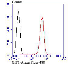
Fig6: Flow cytometric analysis of A549 cells with GIT1 antibody at 1/100 dilution (red) compared with an unlabelled control (cells without incubation with primary antibody; black).| Positive Control | Rat brain tissue, human colon cancer tissue, human pancreas tissue, mouse fallopian tubes tissue, A549. |
|---|---|
| Application Notes | WB:1:500-1:1,000 IHC:1:50-1:200 FC:1:50-1:100 |
| Form | Liquid |
|---|---|
| Storage Instructions | Store at +4℃ after thawing. Aliquot store at -20℃ or -80℃. Avoid repeated freeze / thaw cycles. |
| Storage Buffer | 1*TBS (pH7.4), 1%BSA, 40%Glycerol. Preservative: 0.05% Sodium Azide. |


