Application
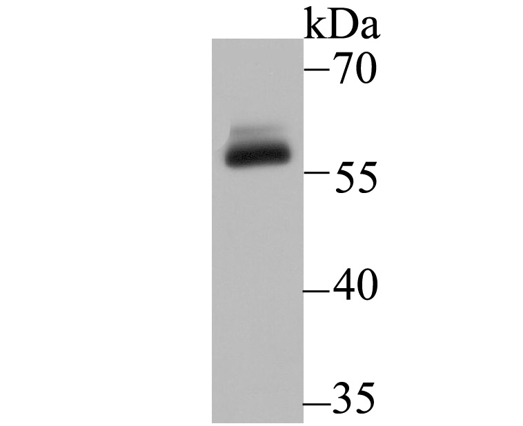
Fig1: Western blot analysis of DKC1 on A431 cell lysate using anti-DKC1 antibody at 1/500 dilution.Application
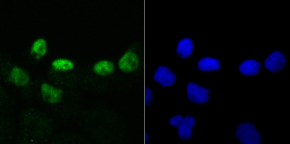
Fig2: ICC staining DKC1 in A431 cells (green). The nuclear counter stain is DAPI (blue). Cells were fixed in paraformaldehyde, permeabilised with 0.25% Triton X100/PBS.Application
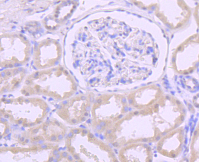
Fig3: Immunohistochemical analysis of paraffin-embedded human kidney tissue using anti-DKC1 antibody. Counter stained with hematoxylin.Application
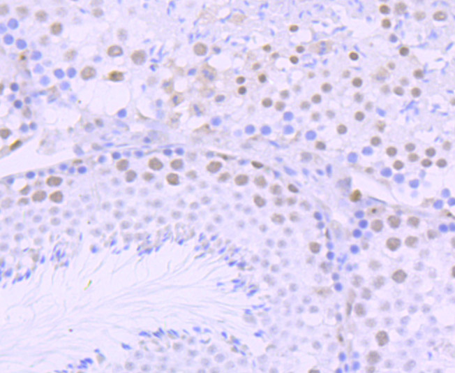
Fig4: Immunohistochemical analysis of paraffin-embedded mouse testis tissue using anti-DKC1 antibody. Counter stained with hematoxylin.Application
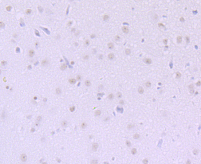
Fig5: Immunohistochemical analysis of paraffin-embedded rat brain tissue using anti-DKC1 antibody. Counter stained with| Product Name | DKC1 [JU34-32] |
|---|---|
| Antibody Type | Primary Antibodies |
| Antigen Alias | CBF5 antibody CBF5 homolog antibody Cbf5p homolog antibody DKC 1 antibody DKC antibody Dkc1 antibody DKC1_HUMAN antibody DKCX antibody Dyskeratosis congenita 1 antibody Dyskeratosis congenita 1 dyskerin antibody Dyskerin antibody H/ACA ribonucleoprotein complex subunit 4 antibody NAP 57 antibody NAP57 antibody NOLA 4 antibody NOLA4 antibody Nopp140 associated protein of 57 kDa antibody Nopp140-associated protein of 57 kDa antibody Nucleolar protein family A member 4 antibody Nucleolar protein NAP57 antibody snoRNP protein DKC1 antibody XAP 101 antibody XAP101 antibody |
| Product description | Isoform 1: Required for ribosome biogenesis and telomere maintenance. Probable catalytic subunit of H/ACA small nucleolar ribonucleoprotein (H/ACA snoRNP) complex, which catalyzes pseudouridylation of rRNA. This involves the isomerization of uridine such that the ribose is subsequently attached to C5, instead of the normal N1. Each rRNA can contain up to 100 pseudouridine ('psi') residues, which may serve to stabilize the conformation of rRNAs. Also required for correct processing or intranuclear trafficking of TERC, the RNA component of the telomerase reverse transcriptase (TERT) holoenzyme. Isoform 3: Promotes cell to cell and cell to substratum adhesion, increases the cell proliferation rate and leads to cytokeratin hyper-expression (when overexpressed in HeLa cells). |
| Immunogen | Recombinant protein |
| Clonality | Monoclonal |
|---|---|
| Isotype | IgG |
| Host Species | Recombinant rabbit |
| Tested Applications | WBICCIFIHC |
| WB:1:500-1:1,000 ICC:1:50-1:200 IHC:1:50-1:100 |
|
| Species Reactivity | HumanMouseRat |
| Concentration | 1mg/ml |
| Alternative Names | CBF5 antibody CBF5 homolog antibody Cbf5p homolog antibody DKC 1 antibody DKC antibody Dkc1 antibody DKC1_HUMAN antibody DKCX antibody Dyskeratosis congenita 1 antibody Dyskeratosis congenita 1 dyskerin antibody Dyskerin antibody H/ACA ribonucleoprotein complex subunit 4 antibody NAP 57 antibody NAP57 antibody NOLA 4 antibody NOLA4 antibody Nopp140 associated protein of 57 kDa antibody Nopp140-associated protein of 57 kDa antibody Nucleolar protein family A member 4 antibody Nucleolar protein NAP57 antibody snoRNP protein DKC1 antibody XAP 101 antibody XAP101 antibody |
|---|---|
| Molecular Weight(MW) | 57 kDa |
| Cellular Localization | Cytoplasm. Nucleus. |
Application
Fig1: Western blot analysis of DKC1 on A431 cell lysate using anti-DKC1 antibody at 1/500 dilution.
Application
Fig2: ICC staining DKC1 in A431 cells (green). The nuclear counter stain is DAPI (blue). Cells were fixed in paraformaldehyde, permeabilised with 0.25% Triton X100/PBS.
Application
Fig3: Immunohistochemical analysis of paraffin-embedded human kidney tissue using anti-DKC1 antibody. Counter stained with hematoxylin.
Application
Fig4: Immunohistochemical analysis of paraffin-embedded mouse testis tissue using anti-DKC1 antibody. Counter stained with hematoxylin.
Application
Fig5: Immunohistochemical analysis of paraffin-embedded rat brain tissue using anti-DKC1 antibody. Counter stained with| Positive Control | A431, rat brain tissue, human kidney tissue, mouse testis tissue. |
|---|---|
| Application Notes | WB:1:500-1:1,000 ICC:1:50-1:200 IHC:1:50-1:100 |
| Form | Liquid |
|---|---|
| Storage Instructions | Store at +4℃ after thawing. Aliquot store at -20℃ or -80℃. Avoid repeated freeze / thaw cycles. |
| Storage Buffer | 1*TBS (pH7.4), 1%BSA, 40%Glycerol. Preservative: 0.05% Sodium Azide. |


