Application
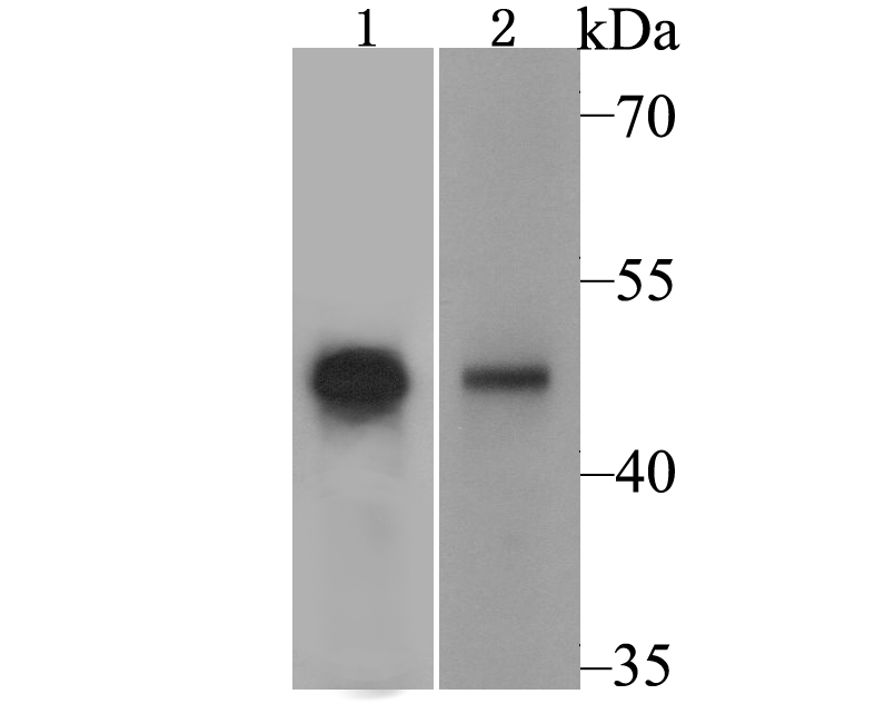
Fig1: Western blot analysis of PPAR gamma on PC-12 cell and human liver tissue lysate using anti-PPAR gamma antibody at 1/500 dilution.Application
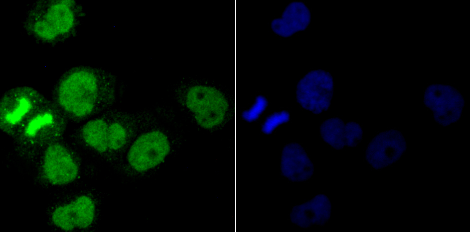
Fig2: ICC staining PPAR gamma in A431 cells (green). The nuclear counter stain is DAPI (blue). Cells were fixed in paraformaldehyde, permeabilised with 0.25% Triton X100/PBS.Application
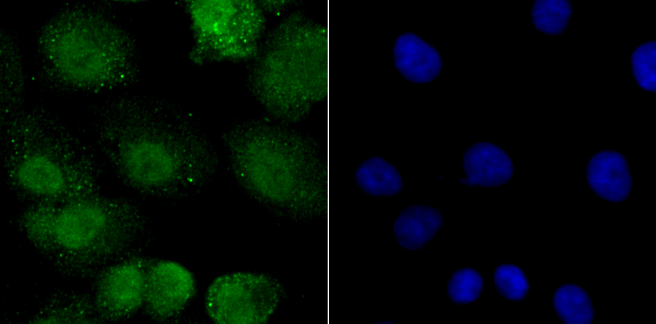
Fig3: ICC staining PPAR gamma in A549 cells (green). The nuclear counter stain is DAPI (blue). Cells were fixed in paraformaldehyde, permeabilised with 0.25% Triton X100/PBS.Application
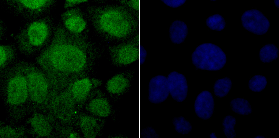
Fig4: ICC staining PPAR gamma in JAR cells (green). The nuclear counter stain is DAPI (blue). Cells were fixed in paraformaldehyde, permeabilised with 0.25% Triton X100/PBS.Application
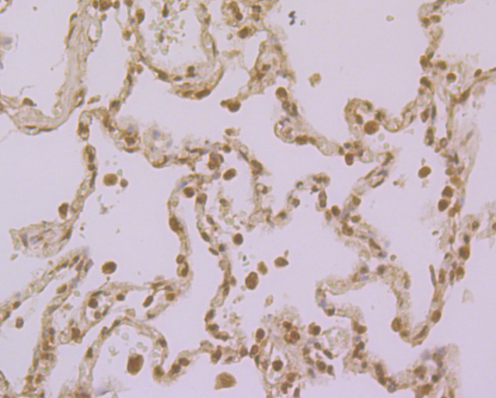
Fig5: Immunohistochemical analysis of paraffin-embedded human lung cancer tissue using anti-PPAR gamma antibody. Counter stained with hematoxylin.Application
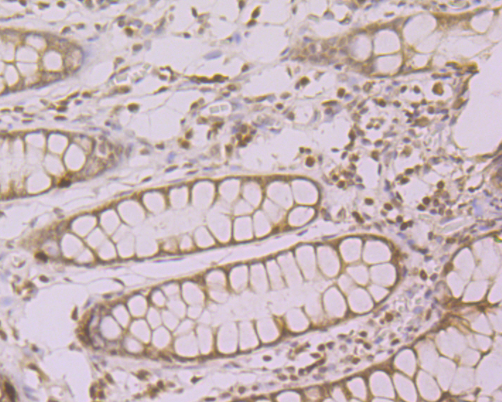
Fig6: Immunohistochemical analysis of paraffin-embedded human colon tissue using anti-PPAR gamma antibody. Counter stained with hematoxylin.Application
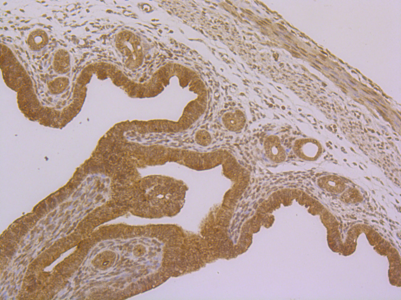
Fig7: Immunohistochemical analysis of paraffin-embedded mouse uterus tissue using anti-PPAR gamma antibody. Counter stained with hematoxylin.Application
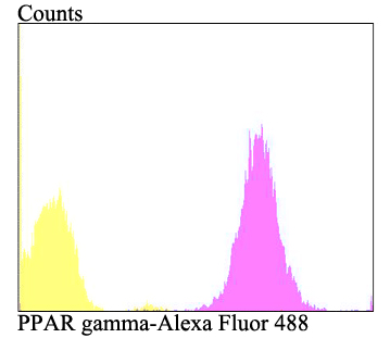
Fig8: Flow cytometric analysis of A431 cells with PPAR gamma antibody at 1/100 dilution (purple) compared with an unlabelled control (cells without incubation with primary antibody; yellow). Alexa Fluor 488-conjugated goat anti-rabbit IgG was used as the secondary antibody.| Product Name | PPAR gamma |
|---|---|
| Antibody Type | Primary Antibodies |
| Antigen Alias | CIMT1 antibody GLM1 antibody NR1C3 antibody Nuclear receptor subfamily 1 group C member 3 antibody OTTHUMP00000185032 antibody OTTHUMP00000185036 antibody Peroxisome proliferator activated nuclear receptor gamma variant 1 antibody Peroxisome proliferator activated receptor gamma 1 antibody Peroxisome Proliferator Activated Receptor gamma antibody Peroxisome proliferator-activated receptor gamma antibody PPAR gamma antibody PPAR-gamma antibody PPARG antibody PPARG_HUMAN antibody PPARG1 antibody PPARG2 antibody PPARgamma antibody |
| Product description | Peroxisome proliferator-activated receptors (PPARs) are members of the nuclear hormone receptor subfamily of transcription factors. PPARs form heterodimers with retinoid X receptors (RXRs). These heterodimers regulate transcription of genes involved in insulin action, adipocyte differentiation, lipid metabolism and inflammation. PPARγ is implicated in numerous diseases including obesity, diabetes, atherosclerosis and cancer. PPARγ activators include prostanoids, fatty acids, thiazolidinediones and N-(2-benzoylphenyl) tyrosine analogues. A key component in adipocyte differentiation and fat-specific gene expression, PPARγ may modulate macrophage functions such as proinflammatory activities, and stimulate oxidized low-density lipoprotein (x-LDL) uptake. A Pro12Ala polymorphism of the PPARγ2 gene has been reported to reduce transactivation activity in vitro. This substitution may affect the immune response to ox-LDL and be associated with type 2 diabetes. In addition, the Pro12Ala variant of the PPARγ2 gene maybe correlated with abdominal obesity in type 2 diabetes. |
| Immunogen | Recombinant protein within human PPAR gamma aa 250-450. |
| Clonality | Polyclonal |
|---|---|
| Isotype | IgG |
| Host Species | Rabbit |
| Tested Applications | WBICCIHCFC |
| WB:1:500 ICC:1:100 IHC:1:50-1:100 FC:1:50-1:100 |
|
| Species Reactivity | HumanMouseRat |
| Concentration | 1mg/ml |
| Alternative Names | CIMT1 antibody GLM1 antibody NR1C3 antibody Nuclear receptor subfamily 1 group C member 3 antibody OTTHUMP00000185032 antibody OTTHUMP00000185036 antibody Peroxisome proliferator activated nuclear receptor gamma variant 1 antibody Peroxisome proliferator activated receptor gamma 1 antibody Peroxisome Proliferator Activated Receptor gamma antibody Peroxisome proliferator-activated receptor gamma antibody PPAR gamma antibody PPAR-gamma antibody PPARG antibody PPARG_HUMAN antibody PPARG1 antibody PPARG2 antibody PPARgamma antibody |
|---|---|
| Molecular Weight(MW) | 57/54/21 kDa |
| Cellular Localization | Nucleus. Cytoplasm. |
| SwissProt ID | P37231 |
|---|
Application
Fig1: Western blot analysis of PPAR gamma on PC-12 cell and human liver tissue lysate using anti-PPAR gamma antibody at 1/500 dilution.
Application
Fig2: ICC staining PPAR gamma in A431 cells (green). The nuclear counter stain is DAPI (blue). Cells were fixed in paraformaldehyde, permeabilised with 0.25% Triton X100/PBS.
Application
Fig3: ICC staining PPAR gamma in A549 cells (green). The nuclear counter stain is DAPI (blue). Cells were fixed in paraformaldehyde, permeabilised with 0.25% Triton X100/PBS.
Application
Fig4: ICC staining PPAR gamma in JAR cells (green). The nuclear counter stain is DAPI (blue). Cells were fixed in paraformaldehyde, permeabilised with 0.25% Triton X100/PBS.
Application
Fig5: Immunohistochemical analysis of paraffin-embedded human lung cancer tissue using anti-PPAR gamma antibody. Counter stained with hematoxylin.
Application
Fig6: Immunohistochemical analysis of paraffin-embedded human colon tissue using anti-PPAR gamma antibody. Counter stained with hematoxylin.
Application
Fig7: Immunohistochemical analysis of paraffin-embedded mouse uterus tissue using anti-PPAR gamma antibody. Counter stained with hematoxylin.
Application
Fig8: Flow cytometric analysis of A431 cells with PPAR gamma antibody at 1/100 dilution (purple) compared with an unlabelled control (cells without incubation with primary antibody; yellow). Alexa Fluor 488-conjugated goat anti-rabbit IgG was used as the secondary antibody.| Positive Control | PC-12, human liver tissue lysate, A431, A549, JAR, human lung cancer tissue, human colon tissue, mouse uterus tissue. |
|---|---|
| Application Notes | WB:1:500 ICC:1:100 IHC:1:50-1:100 FC:1:50-1:100 |
| Form | Liquid |
|---|---|
| Storage Instructions | Store at +4℃ after thawing. Aliquot store at -20℃. Avoid repeated freeze / thaw cycles. |
| Storage Buffer | 1*TBS (pH7.4), 0.5%BSA, 50%Glycerol. Preservative: 0.05% Sodium Azide. |


