Application
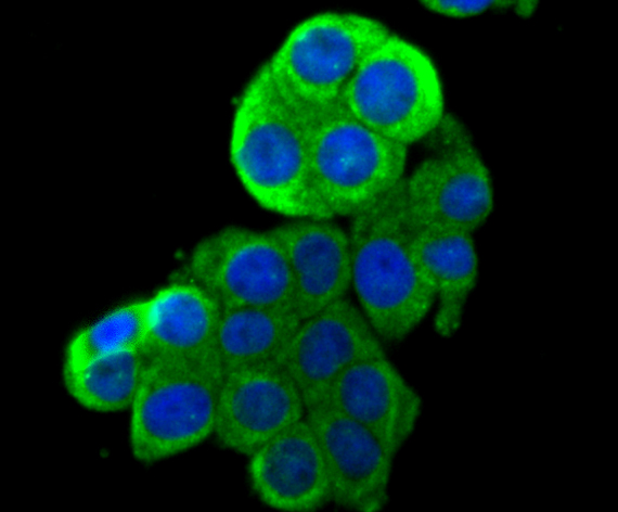
Fig1: ICC staining HKDC1 in LOVO cells (green). The nuclear counter stain is DAPI (blue). Cells were fixed in paraformaldehyde, permeabilised with 0.25% Triton X100/PBS.Application
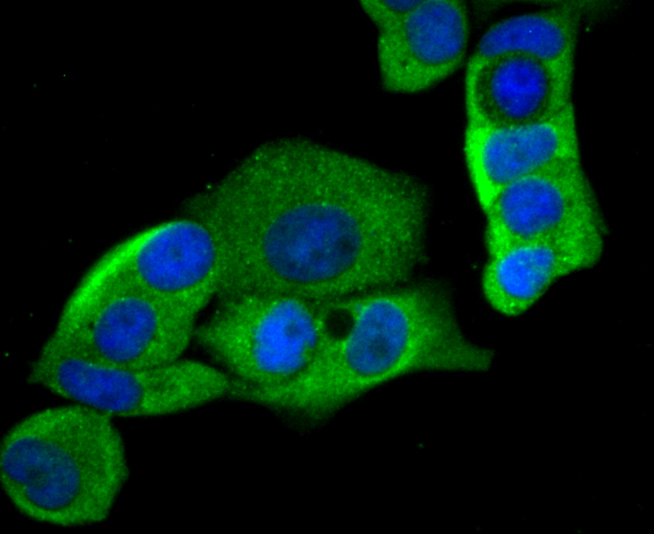
Fig2: ICC staining HKDC1 in MCF-7 cells (green). The nuclear counter stain is DAPI (blue). Cells were fixed in paraformaldehyde, permeabilised with 0.25% Triton X100/PBS.Application
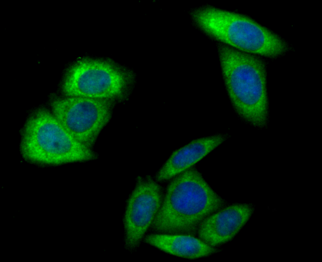
Fig3: ICC staining HKDC1 in SiHa cells (green). The nuclear counter stain is DAPI (blue). Cells were fixed in paraformaldehyde, permeabilised with 0.25% Triton X100/PBS.Application
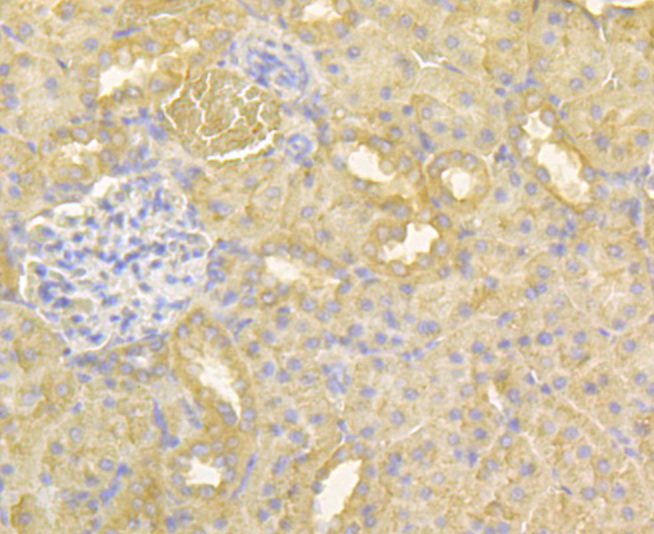
Fig4: Immunohistochemical analysis of paraffin-embedded rat kidney tissue using anti-HKDC1 antibody. Counter stained with hematoxylin.Application
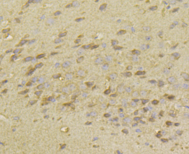
Fig5: Immunohistochemical analysis of paraffin-embedded mouse brain tissue using anti-HKDC1 antibody. Counter stained with hematoxylin.Application
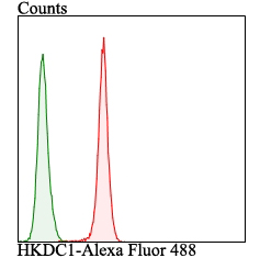
Fig6: Flow cytometric analysis of LOVO cells with HKDC1 antibody at 1/100 dilution (red) compared with an unlabelled control (cells without incubation with primary antibody; green). Alexa Fluor 488-conjugated goat anti-rabbit IgG was used as the secondary antibody.| Product Name | HKDC1 |
|---|---|
| Antibody Type | Primary Antibodies |
| Antigen Alias | BC016235 antibody Hexokinase domain containing 1 antibody Hexokinase domain-containing protein 1 antibody Hkdc1 antibody HKDC1_HUMAN antibody Putative hexokinase HKDC1 antibody |
| Product description | As the recently identified fifth isoform of hexokinase, HKDC1 catalyzes the rate-limiting and first obligatory step of glucose metabolism, which is the ATP-dependent phosphorylation of glucose to G6P. Though its particular biological function remains unclear, HKDC1 has been suggested to play a more major role in glucose metabolism during pregnancy, as the mother would need to provide enough energy for both herself and the fetus. HKDC1 is ubiquitously expressed, with the highest levels of expression in pharynx, thymus, colon, esophagus, and eye tissue. |
| Immunogen | Recombinant protein within human HKDC1 260-460aa. |
| Clonality | Polyclonal |
|---|---|
| Isotype | IgG |
| Host Species | Rabbit |
| Tested Applications | WBICCIHCFC |
| ICC:1:50-1:200 IHC:1:50-1:200 FC:1:50-1:100 WB:1:500 |
|
| Species Reactivity | HumanMouseRat |
| Concentration | 1mg/ml |
| Alternative Names | BC016235 antibody Hexokinase domain containing 1 antibody Hexokinase domain-containing protein 1 antibody Hkdc1 antibody HKDC1_HUMAN antibody Putative hexokinase HKDC1 antibody |
|---|---|
| Molecular Weight(MW) | 82/90/102 kDa |
| Cellular Localization | Cytosol. Mitochondrion. |
Application
Fig1: ICC staining HKDC1 in LOVO cells (green). The nuclear counter stain is DAPI (blue). Cells were fixed in paraformaldehyde, permeabilised with 0.25% Triton X100/PBS.
Application
Fig2: ICC staining HKDC1 in MCF-7 cells (green). The nuclear counter stain is DAPI (blue). Cells were fixed in paraformaldehyde, permeabilised with 0.25% Triton X100/PBS.
Application
Fig3: ICC staining HKDC1 in SiHa cells (green). The nuclear counter stain is DAPI (blue). Cells were fixed in paraformaldehyde, permeabilised with 0.25% Triton X100/PBS.
Application
Fig4: Immunohistochemical analysis of paraffin-embedded rat kidney tissue using anti-HKDC1 antibody. Counter stained with hematoxylin.
Application
Fig5: Immunohistochemical analysis of paraffin-embedded mouse brain tissue using anti-HKDC1 antibody. Counter stained with hematoxylin.
Application
Fig6: Flow cytometric analysis of LOVO cells with HKDC1 antibody at 1/100 dilution (red) compared with an unlabelled control (cells without incubation with primary antibody; green). Alexa Fluor 488-conjugated goat anti-rabbit IgG was used as the secondary antibody.| Positive Control | LOVO, MCF-7, SiHa, rat kidney tissue, mouse brain tissue. |
|---|---|
| Application Notes | ICC:1:50-1:200 IHC:1:50-1:200 FC:1:50-1:100 WB:1:500 |
| Form | Liquid |
|---|---|
| Storage Instructions | Store at +4℃ after thawing. Aliquot store at -20℃. Avoid repeated freeze / thaw cycles. |
| Storage Buffer | 1*TBS (pH7.4), 0.5%BSA, 50%Glycerol. Preservative: 0.05% Sodium Azide. |


