Application
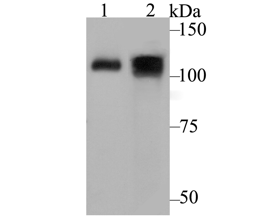
Fig1: Western blot analysis of ATP2A1 on different lysates using anti-ATP2A1 antibody at 1/500 dilution. Positive control: Lane1: Mouse skeletal muscle tissue Lane2: Human skeletal muscle tissueApplication
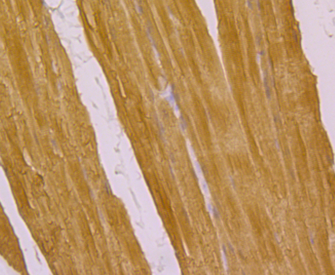
Fig2: Immunohistochemical analysis of paraffin-embedded rat skeletal muscle tissue using anti-ATP2A1 antibody. Counter stained with hematoxylin.Application
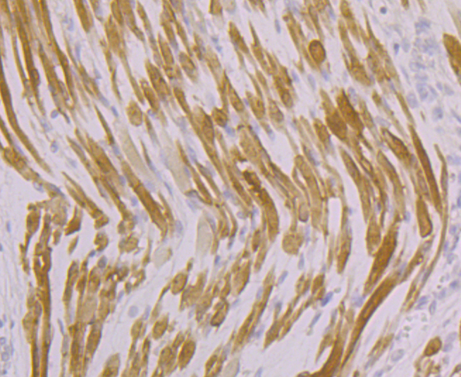
Fig3: Immunohistochemical analysis of paraffin-embedded human fetal skeletal muscle tissue using anti-ATP2A1 antibody. Counter stained with hematoxylin.Application
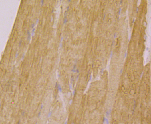
Fig4: Immunohistochemical analysis of paraffin-embedded mouse skeletal muscle tissue using anti-ATP2A1 antibody. Counter stained with hematoxylin.Application
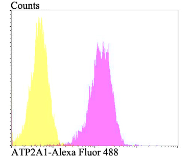
Fig5: Flow cytometric analysis of 293T cells with ATP2A1 antibody at 1/100 dilution (fuchsia) compared with an unlabelled control (cells without incubation with primary antibody; yellow). Alexa Fluor 488-conjugated goat anti-rabbit IgG was used as the secondary antibody.| Product Name | ATP2A1 |
|---|---|
| Antibody Type | Primary Antibodies |
| Antigen Alias | fast twitch skeletal muscle isoform antibody AT2A1_HUMAN antibody ATP2A antibody ATP2A1 antibody ATPase Ca++ transporting cardiac muscle fast twitch 1 antibody ATPase Ca++ transporting fast twitch 1 antibody ATPase, Ca(2+)-transporting fast twitch 1 antibody Calcium pump 1 antibody Calcium transporting ATPase sarcoplasmic reticulum type fast twitch skeletal muscle isoform antibody Calcium-transporting ATPase sarcoplasmic reticulum type antibody EC 3.6.3.8 antibody Endoplasmic reticulum class 1/2 Ca(2+) ATPase antibody Fast skeletal muscle SR calcium ATPase antibody OTTHUMP00000162561 antibody OTTHUMP00000162562 antibody Sarcoendoplasmic reticulum calcium ATPase antibody Sarcoplasmic reticulum Ca(2+)-ATPase 1 antibody Sarcoplasmic/endoplasmic reticulum calcium ATPase 1 antibody SERCA 1 antibody SERCA1 antibody SERCA1 truncated isoform, included antibody SR Ca(2+) ATPase 1 antibody SR Ca(2+)-ATPase 1 antibody |
| Product description | Key regulator of striated muscle performance by acting as the major Ca2+ ATPase responsible for the reuptake of cytosolic Ca2+ into the sarcoplasmic reticulum. Catalyzes the hydrolysis of ATP coupled with the translocation of calcium from the cytosol to the sarcoplasmic reticulum lumen. Contributes to calcium sequestration involved in muscular excitation/contraction. |
| Immunogen | Synthetis peptide within rat ATP2A1 aa 20-50. |
| Clonality | Polyclonal |
|---|---|
| Isotype | IgG |
| Host Species | Rabbit |
| Tested Applications | WBIHCFC |
| WB:1:500-1:1,000 IHC:1:50-1:200 FC:1:50-1:100 |
|
| Species Reactivity | HumanMouseRat |
| Concentration | 1mg/ml |
| Alternative Names | fast twitch skeletal muscle isoform antibody
AT2A1_HUMAN antibody
ATP2A antibody
ATP2A1 antibody
ATPase Ca++ transporting cardiac muscle fast twitch 1 antibody
ATPase Ca++ transporting fast twitch 1 antibody
ATPase Ca(2+)-transporting fast twitch 1 antibody Calcium pump 1 antibody Calcium transporting ATPase sarcoplasmic reticulum type fast twitch skeletal muscle isoform antibody Calcium-transporting ATPase sarcoplasmic reticulum type antibody EC 3.6.3.8 antibody Endoplasmic reticulum class 1/2 Ca(2+) ATPase antibody Fast skeletal muscle SR calcium ATPase antibody OTTHUMP00000162561 antibody OTTHUMP00000162562 antibody Sarcoendoplasmic reticulum calcium ATPase antibody Sarcoplasmic reticulum Ca(2+)-ATPase 1 antibody Sarcoplasmic/endoplasmic reticulum calcium ATPase 1 antibody SERCA 1 antibody SERCA1 antibody SERCA1 truncated isoform included antibody SR Ca(2+) ATPase 1 antibody SR Ca(2+)-ATPase 1 antibody |
|---|---|
| Molecular Weight(MW) | 110 kDa |
| Cellular Localization | Endoplasmic reticulum membrane. Sarcoplasmic reticulum membrane. |
Application
Fig1: Western blot analysis of ATP2A1 on different lysates using anti-ATP2A1 antibody at 1/500 dilution. Positive control: Lane1: Mouse skeletal muscle tissue Lane2: Human skeletal muscle tissue
Application
Fig2: Immunohistochemical analysis of paraffin-embedded rat skeletal muscle tissue using anti-ATP2A1 antibody. Counter stained with hematoxylin.
Application
Fig3: Immunohistochemical analysis of paraffin-embedded human fetal skeletal muscle tissue using anti-ATP2A1 antibody. Counter stained with hematoxylin.
Application
Fig4: Immunohistochemical analysis of paraffin-embedded mouse skeletal muscle tissue using anti-ATP2A1 antibody. Counter stained with hematoxylin.
Application
Fig5: Flow cytometric analysis of 293T cells with ATP2A1 antibody at 1/100 dilution (fuchsia) compared with an unlabelled control (cells without incubation with primary antibody; yellow). Alexa Fluor 488-conjugated goat anti-rabbit IgG was used as the secondary antibody.| Positive Control | Human skeletal muscle tissue, mouse skeletal muscle tissue, rat skeletal muscle tissue, 293T. |
|---|---|
| Application Notes | WB:1:500-1:1,000 IHC:1:50-1:200 FC:1:50-1:100 |
| Form | Liquid |
|---|---|
| Storage Instructions | Store at +4℃ after thawing. Aliquot store at -20℃. Avoid repeated freeze / thaw cycles. |
| Storage Buffer | 1*TBS (pH7.4), 0.5%BSA, 50%Glycerol. Preservative: 0.05% Sodium Azide. |


