Application
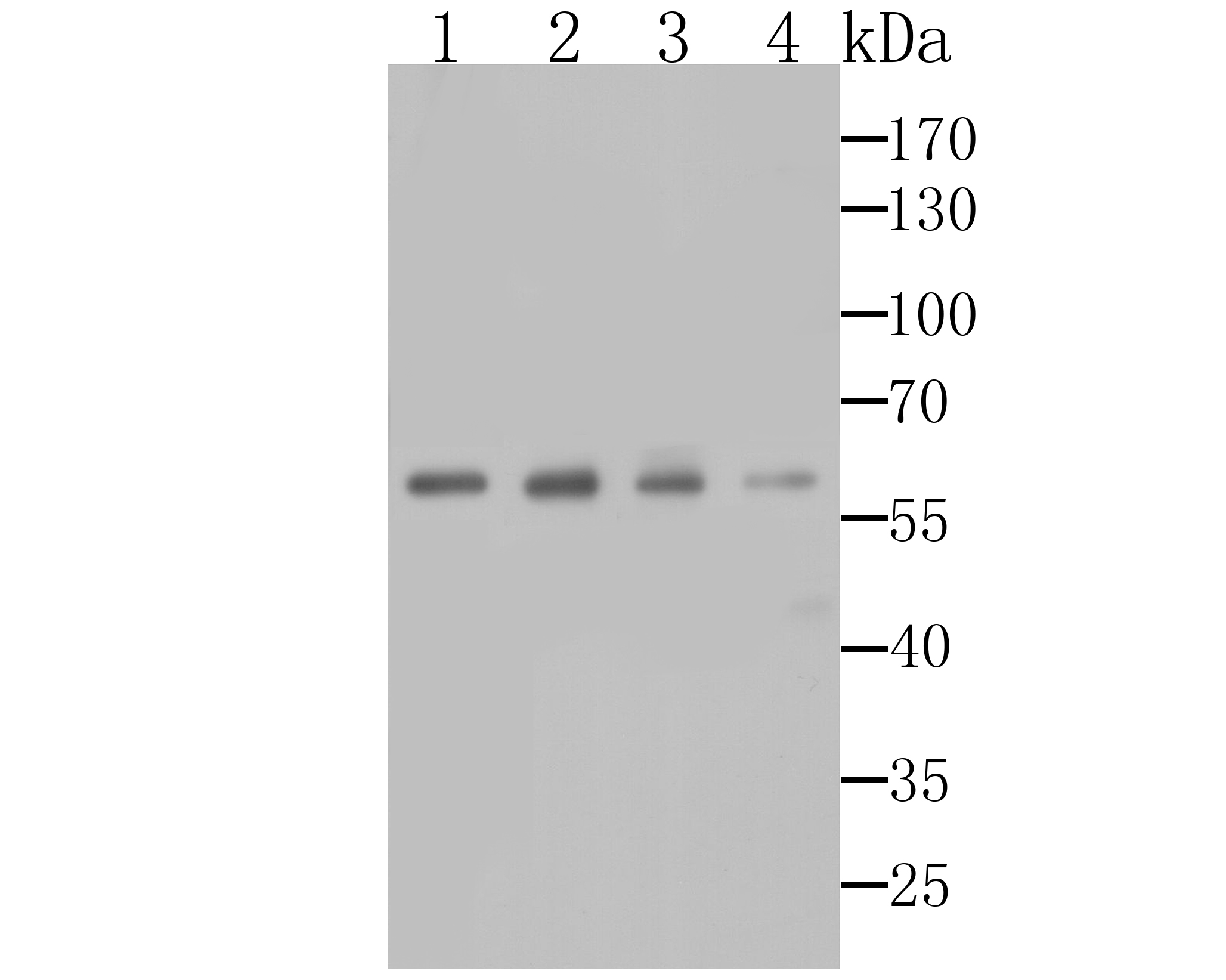
Fig1: Western blot analysis of AMBP on different cell lysates using anti-AMBP antibody at 1/1,000 dilution. Positive control: Lane 1: HepG2 Lane 2: Human liver Lane 3: Mouse liver Lane 4: Mouse brainApplication
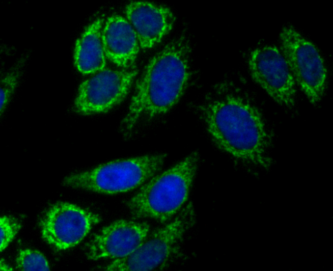
Fig2: ICC staining AMBP in HepG2 cells (green). The nuclear counter stain is DAPI (blue). Cells were fixed in paraformaldehyde, permeabilised with 0.25% Triton X100/PBS.Application
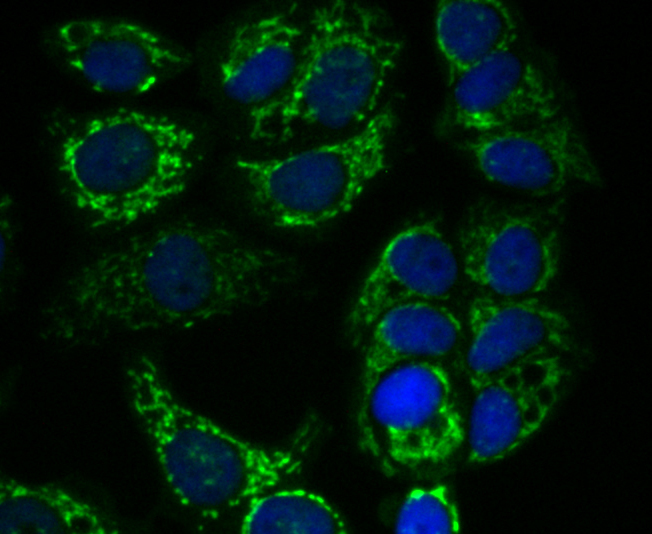
Fig3: ICC staining AMBP in MCF-7 cells (green). The nuclear counter stain is DAPI (blue). Cells were fixed in paraformaldehyde, permeabilised with 0.25% Triton X100/PBS.Application
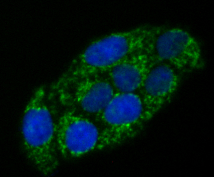
Fig4: ICC staining AMBP in Hela cells (green). The nuclear counter stain is DAPI (blue). Cells were fixed in paraformaldehyde, permeabilised with 0.25% Triton X100/PBS.Application
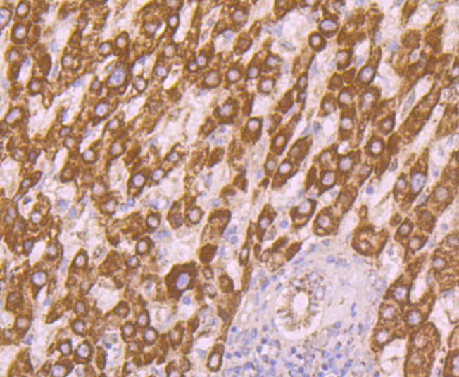
Fig5: Immunohistochemical analysis of paraffin-embedded human liver cancer tissue using anti-AMBP antibody. Counter stained with hematoxylin.Application
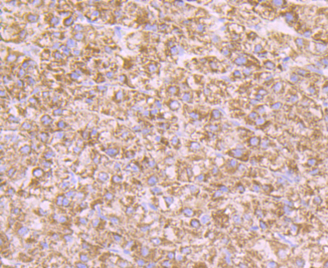
Fig6: Immunohistochemical analysis of paraffin-embedded human liver tissue using anti-AMBP antibody. Counter stained with hematoxylin.Application
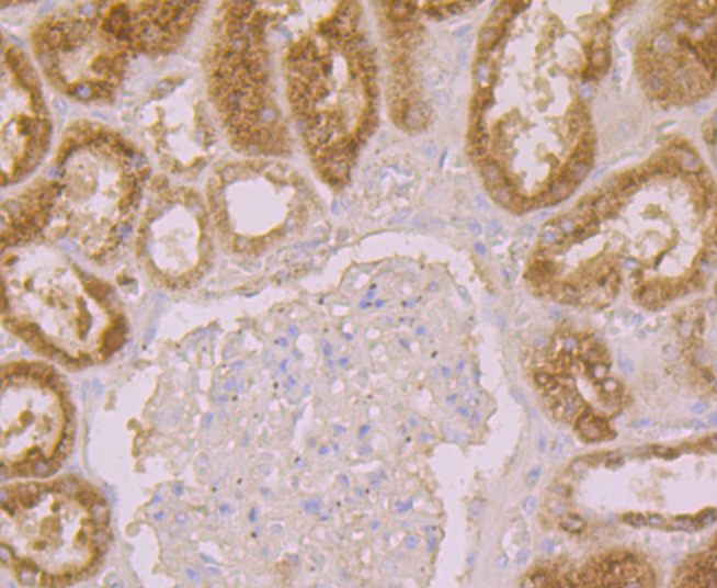
Fig7: Immunohistochemical analysis of paraffin-embedded human kidney tissue using anti-AMBP antibody. Counter stained with hematoxylin.Application
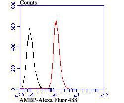
Fig8: Flow cytometric analysis of HepG2 cells with AMBP antibody at 1/100 dilution (red) compared with an unlabelled control (cells without incubation with primary antibody; black). Alexa Fluor 488-conjugated goat anti-rabbit IgG was used as the secondary antibody.| Product Name | AMBP |
|---|---|
| Antibody Type | Primary Antibodies |
| Antigen Alias | Alpha 1 microglobulin/bikunin precursor antibody Alpha-1 microglycoprotein antibody AMBP antibody AMBP_HUMAN antibody Bikunin antibody Complex-forming glycoprotein heterogeneous in charge antibody EDC1 antibody Growth inhibiting protein 19 antibody HI 30 antibody HI-30 antibody HI30 antibody IATIL antibody Inter alpha trypsin inhibitor light chain antibody ITI antibody ITI LC antibody ITI-LC antibody ITILC antibody Protein HC antibody Trypstatin antibody Uronic-acid-rich protein antibody UTI antibody |
| Product description | The AMBP (α-1-Microglobulin/bikunin precursor) gene encodes a protein precursor, known as AMBP, that is cleaved to produce two distinct proteins, designated a-1-Microglobulin and Bikunin. α-1-Microglobulin, also known as protein HC, is a member of the lipocalin superfamily and is secreted mainly in plasma, urine and cerebrospinal fluid. Thought to have reductase/dehydrogenase activity, α-1-Microglobulin exhibits immunosuppressive properties, such as cytokine secretion and inhibition of antigen-induced lymphocyte cell proliferation, and may be involved in the reduction of biological pro-oxidants. The second protein cleavage product, designated Bikunin and also known as inter-α-trypsin inhibitor light chain, ITI-LC or urinary trypsin inhibitor, is a widely expressed protein that is stored in the granules of human connective tissue mast cells. One of many proteins in the Kunitz-type protease inhibitor family, Bikunin prevents autodigestion by exocrine enzymes, such as trypsinogen and chymo-trypsinogen, and plays a role in the antiinflammatory/antiproteinase immune response. Unlike α-1-Microglobulin, Bikunin is implicated in the pathogenesis of a number of renal diseases, such as urolithiasis. |
| Immunogen | Recombinant protein with Human AMBP aa 120-320. |
| Clonality | Polyclonal |
|---|---|
| Isotype | IgG |
| Host Species | Rabbit |
| Tested Applications | WBICCIHCFC |
| WB:1:500-1:1,000 ICC:1:50-1:200 IHC:1:50-1:200 FC:1:50-1:100 |
|
| Species Reactivity | HumanMouse |
| Concentration | 1mg/ml |
| Alternative Names | Alpha 1 microglobulin/bikunin precursor antibody Alpha-1 microglycoprotein antibody AMBP antibody AMBP_HUMAN antibody Bikunin antibody Complex-forming glycoprotein heterogeneous in charge antibody EDC1 antibody Growth inhibiting protein 19 antibody HI 30 antibody HI-30 antibody HI30 antibody IATIL antibody Inter alpha trypsin inhibitor light chain antibody ITI antibody ITI LC antibody ITI-LC antibody ITILC antibody Protein HC antibody Trypstatin antibody Uronic-acid-rich protein antibody UTI antibody |
|---|---|
| Molecular Weight(MW) | 39 kDa (Predicted band size) |
| Cellular Localization | Secreted. |
| SwissProt ID | P02760 |
|---|
Application
Fig1: Western blot analysis of AMBP on different cell lysates using anti-AMBP antibody at 1/1,000 dilution. Positive control: Lane 1: HepG2 Lane 2: Human liver Lane 3: Mouse liver Lane 4: Mouse brain
Application
Fig2: ICC staining AMBP in HepG2 cells (green). The nuclear counter stain is DAPI (blue). Cells were fixed in paraformaldehyde, permeabilised with 0.25% Triton X100/PBS.
Application
Fig3: ICC staining AMBP in MCF-7 cells (green). The nuclear counter stain is DAPI (blue). Cells were fixed in paraformaldehyde, permeabilised with 0.25% Triton X100/PBS.
Application
Fig4: ICC staining AMBP in Hela cells (green). The nuclear counter stain is DAPI (blue). Cells were fixed in paraformaldehyde, permeabilised with 0.25% Triton X100/PBS.
Application
Fig5: Immunohistochemical analysis of paraffin-embedded human liver cancer tissue using anti-AMBP antibody. Counter stained with hematoxylin.
Application
Fig6: Immunohistochemical analysis of paraffin-embedded human liver tissue using anti-AMBP antibody. Counter stained with hematoxylin.
Application
Fig7: Immunohistochemical analysis of paraffin-embedded human kidney tissue using anti-AMBP antibody. Counter stained with hematoxylin.
Application
Fig8: Flow cytometric analysis of HepG2 cells with AMBP antibody at 1/100 dilution (red) compared with an unlabelled control (cells without incubation with primary antibody; black). Alexa Fluor 488-conjugated goat anti-rabbit IgG was used as the secondary antibody.| Positive Control | Human liver, Mouse liver, Mouse brain, Hela, HepG2, MCF-7, human liver tissue, human liver cancer tissue, human kidney tissue. |
|---|---|
| Application Notes | WB:1:500-1:1,000 ICC:1:50-1:200 IHC:1:50-1:200 FC:1:50-1:100 |
| Form | Liquid |
|---|---|
| Storage Instructions | Store at +4℃ after thawing. Aliquot store at -20℃. Avoid repeated freeze / thaw cycles. |
| Storage Buffer | 1*TBS (pH7.4), 0.5%BSA, 50%Glycerol. Preservative: 0.05% Sodium Azide. |


