Application
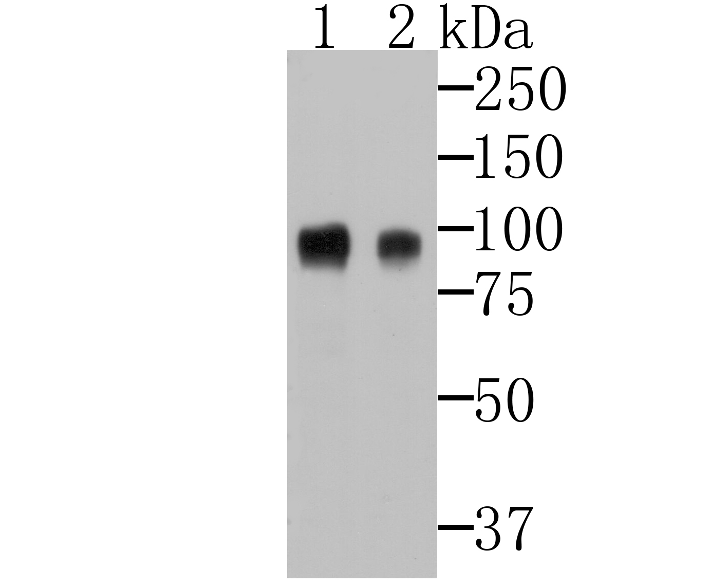
Fig1: Western blot analysis of CD276 on 293 and 293T cell lysates using anti-CD276 antibody at 1/500 dilution.Application
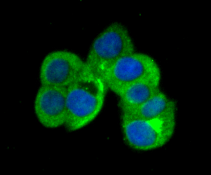
Fig2: ICC staining CD276 in LOVO cells (green). The nuclear counter stain is DAPI (blue). Cells were fixed in paraformaldehyde, permeabilised with 0.25% Triton X100/PBS.Application
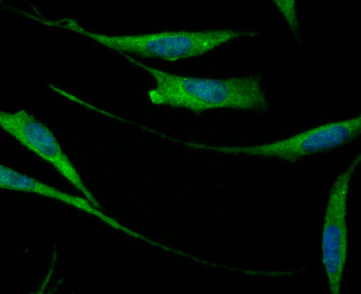
Fig3: ICC staining CD276 in SH-SY-5Y cells (green). The nuclear counter stain is DAPI (blue). Cells were fixed in paraformaldehyde, permeabilised with 0.25% Triton X100/PBS.Application
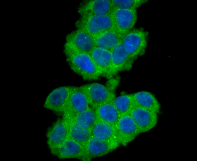
Fig4: ICC staining CD276 in 293T cells (green). The nuclear counter stain is DAPI (blue). Cells were fixed in paraformaldehyde, permeabilised with 0.25% Triton X100/PBS.Application
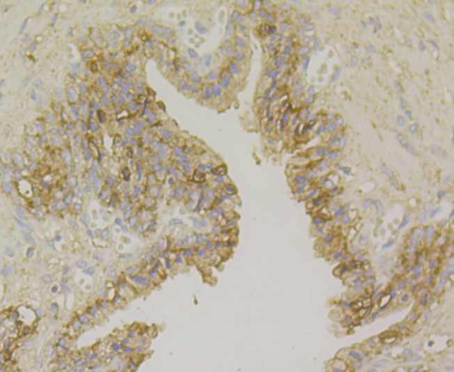
Fig5: Immunohistochemical analysis of paraffin-embedded human prostate tissue using anti-CD276 antibody. Counter stained with hematoxylin.Application
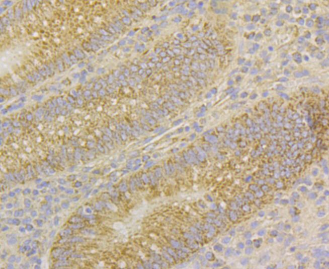
Fig6: Immunohistochemical analysis of paraffin-embedded human appendix tissue using anti-CD276 antibody. Counter stained with hematoxylin.Application
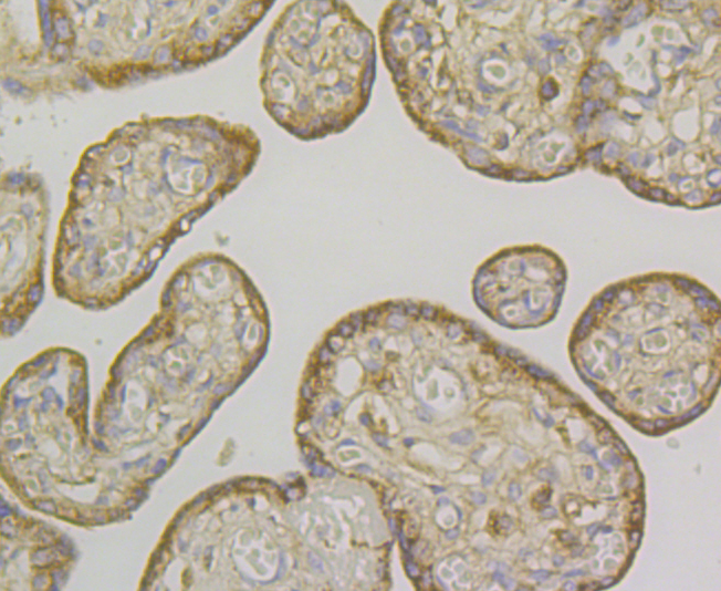
Fig7: Immunohistochemical analysis of paraffin-embedded human placenta tissue using anti-CD276 antibody. Counter stained with hematoxylin.Application
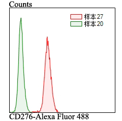
Fig8: Flow cytometric analysis of LOVO cells with CD276 antibody at 1/100 dilution (red) compared with an unlabelled control (cells without incubation with primary antibody; green). Alexa Fluor 488-conjugated goat anti-rabbit IgG was used as the secondary antibody.| Product Name | CD276 |
|---|---|
| Antibody Type | Primary Antibodies |
| Antigen Alias | 4Ig B7 H3 antibody 4Ig-B7-H3 antibody AU016588 antibody B7 H3 antibody B7 homolog 3 antibody B7-H3 antibody B7H3 antibody B7RP-2 antibody CD_antigen=CD276 antibody CD276 antibody CD276 antigen antibody CD276 molecule antibody CD276_HUMAN antibody Costimulatory molecule antibody Flags: Precursor antibody PSEC0249 antibody UNQ309/PRO352 antibody |
| Product description | May participate in the regulation of T-cell-mediated immune response. May play a protective role in tumor cells by inhibiting natural-killer mediated cell lysis as well as a role of marker for detection of neuroblastoma cells. May be involved in the development of acute and chronic transplant rejection and in the regulation of lymphocytic activity at mucosal surfaces. Could also play a key role in providing the placenta and fetus with a suitable immunological environment throughout pregnancy. Both isoform 1 and isoform 2 appear to be redundant in their ability to modulate CD4 T-cell responses. Isoform 2 is shown to enhance the induction of cytotoxic T-cells and selectively stimulates interferon gamma production in the presence of T-cell receptor signaling. |
| Immunogen | Recombinant protein within human CD276 280-500 aa. |
| Clonality | Polyclonal |
|---|---|
| Isotype | IgG |
| Host Species | Rabbit |
| Tested Applications | WBICCIHCFC |
| WB:1:500-1:2,000 ICC:1:50-1:200 IHC:1:50-1:200 FC:1:50-1:100 |
|
| Species Reactivity | Human |
| Concentration | 1mg/ml |
| Alternative Names | 4Ig B7 H3 antibody 4Ig-B7-H3 antibody AU016588 antibody B7 H3 antibody B7 homolog 3 antibody B7-H3 antibody B7H3 antibody B7RP-2 antibody CD_antigen=CD276 antibody CD276 antibody CD276 antigen antibody CD276 molecule antibody CD276_HUMAN antibody Costimulatory molecule antibody Flags: Precursor antibody PSEC0249 antibody UNQ309/PRO352 antibody |
|---|---|
| Molecular Weight(MW) | 57 kDa (Predicted band size) |
| Cellular Localization | Membrane. |
| SwissProt ID | Q5ZPR3 |
|---|
Application
Fig1: Western blot analysis of CD276 on 293 and 293T cell lysates using anti-CD276 antibody at 1/500 dilution.
Application
Fig2: ICC staining CD276 in LOVO cells (green). The nuclear counter stain is DAPI (blue). Cells were fixed in paraformaldehyde, permeabilised with 0.25% Triton X100/PBS.
Application
Fig3: ICC staining CD276 in SH-SY-5Y cells (green). The nuclear counter stain is DAPI (blue). Cells were fixed in paraformaldehyde, permeabilised with 0.25% Triton X100/PBS.
Application
Fig4: ICC staining CD276 in 293T cells (green). The nuclear counter stain is DAPI (blue). Cells were fixed in paraformaldehyde, permeabilised with 0.25% Triton X100/PBS.
Application
Fig5: Immunohistochemical analysis of paraffin-embedded human prostate tissue using anti-CD276 antibody. Counter stained with hematoxylin.
Application
Fig6: Immunohistochemical analysis of paraffin-embedded human appendix tissue using anti-CD276 antibody. Counter stained with hematoxylin.
Application
Fig7: Immunohistochemical analysis of paraffin-embedded human placenta tissue using anti-CD276 antibody. Counter stained with hematoxylin.
Application
Fig8: Flow cytometric analysis of LOVO cells with CD276 antibody at 1/100 dilution (red) compared with an unlabelled control (cells without incubation with primary antibody; green). Alexa Fluor 488-conjugated goat anti-rabbit IgG was used as the secondary antibody.| Positive Control | 293T, LOVO, SH-SY-5Y, human appendix tissue, human prostate tissue, human placenta tissue. |
|---|---|
| Application Notes | WB:1:500-1:2,000 ICC:1:50-1:200 IHC:1:50-1:200 FC:1:50-1:100 |
| Form | Liquid |
|---|---|
| Storage Instructions | Store at +4℃ after thawing. Aliquot store at -20℃. Avoid repeated freeze / thaw cycles. |
| Storage Buffer | 1*TBS (pH7.4), 0.5%BSA, 50%Glycerol. Preservative: 0.05% Sodium Azide. |


