Application
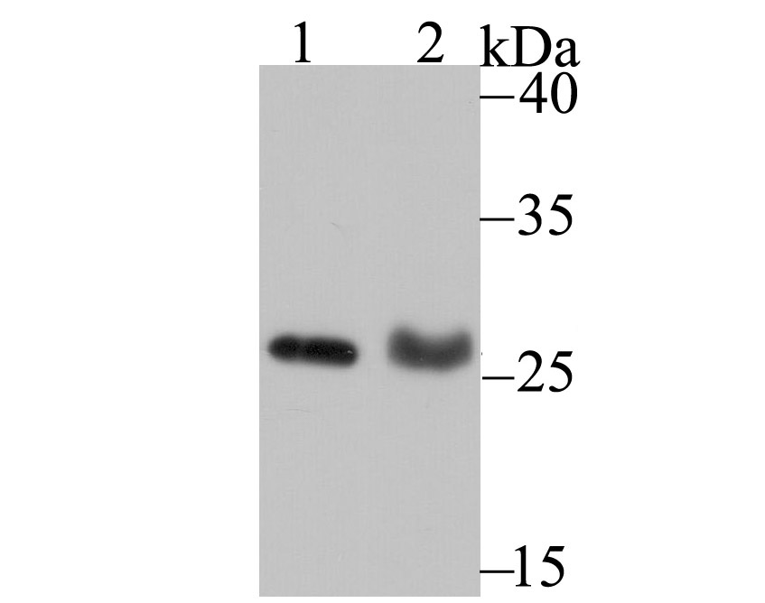
Fig1: Western blot analysis of PSME1 on MCF-7 cell (1) and rat lung tissue (2) lysate using anti-PSME1 antibody at 1/500 dilution.Application
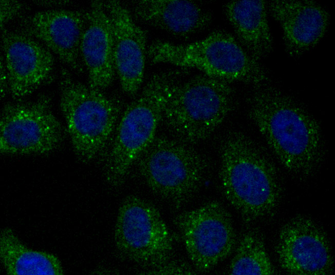
Fig2: ICC staining PSME1 in HepG2 cells (green). The nuclear counter stain is DAPI (blue). Cells were fixed in paraformaldehyde, permeabilised with 0.25% Triton X100/PBS.Application
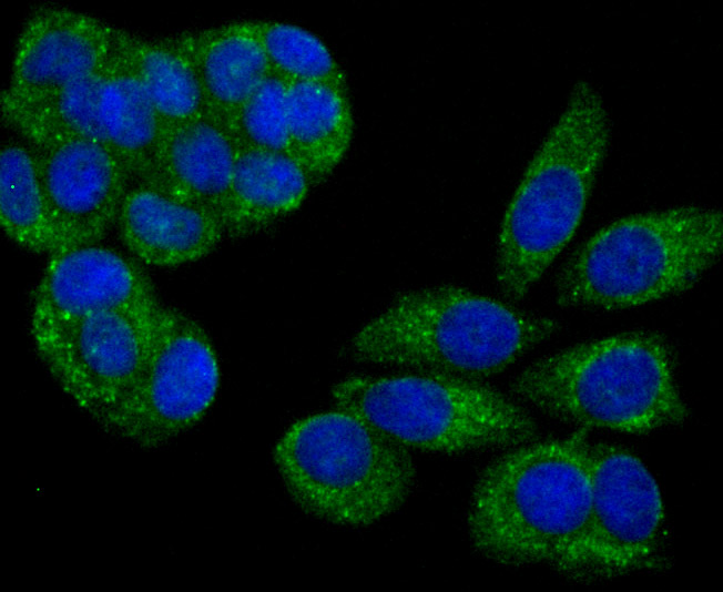
Fig3: ICC staining PSME1 in MCF-7 cells (green). The nuclear counter stain is DAPI (blue). Cells were fixed in paraformaldehyde, permeabilised with 0.25% Triton X100/PBS.Application
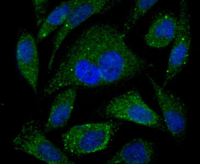
Fig4: ICC staining PSME1 in PC-3M cells (green). The nuclear counter stain is DAPI (blue). Cells were fixed in paraformaldehyde, permeabilised with 0.25% Triton X100/PBS.Application
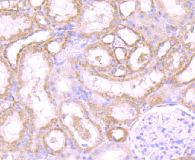
Fig5: Immunohistochemical analysis of paraffin-embedded rat kidney tissue using anti-PSME1 antibody. Counter stained with hematoxylin.Application
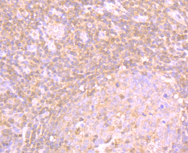
Fig6: Immunohistochemical analysis of paraffin-embedded human tonsil tissue using anti-PSME1 antibody. Counter stained with hematoxylin.Application
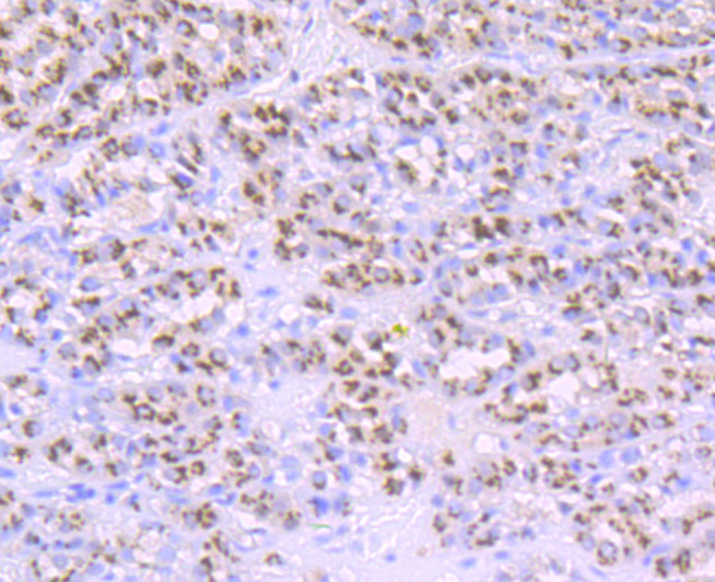
Fig7: Immunohistochemical analysis of paraffin-embedded human liver cancer tissue using anti-PSME1 antibody. Counter stained with hematoxylin.Application
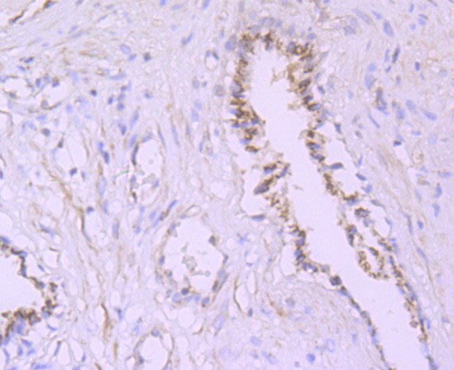
Fig8: Immunohistochemical analysis of paraffin-embedded human prostate tissue using anti-PSME1 antibody. Counter stained with hematoxylin.Application
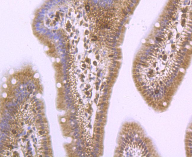
Fig9: Immunohistochemical analysis of paraffin-embedded mouse small intestine tissue using anti-PSME1 antibody. Counter stained with hematoxylin.| Product Name | PSME1 [JU60-33] |
|---|---|
| Antibody Type | Primary Antibodies |
| Antigen Alias | 11S regulator complex alpha subunit antibody 11S regulator complex subunit alpha antibody 29 kD MCP activator subunit antibody 29kD MCP activator subunit antibody Activator of multicatalytic protease subunit 1 antibody IFI5111 antibody IGUP I-5111 antibody Interferon gamma IEF SSP 5111 antibody Interferon gamma inducible protein 5111 antibody Interferon gamma up regulated I 5111 protein antibody Interferon gamma up-regulated I-5111 protein antibody MGC8628 antibody PA28a antibody PA28alpha antibody Proteasome (prosome, macropain) activator subunit 1 (PA28 alpha) antibody Proteasome activator 28 subunit alpha antibody Proteasome activator complex subunit 1 antibody Proteasome activator subunit 1 antibody PSME1 antibody PSME1_HUMAN antibody REG-alpha antibody REGalpha antibody |
| Product description | Implicated in immunoproteasome assembly and required for efficient antigen processing. The PA28 activator complex enhances the generation of class I binding peptides by altering the cleavage pattern of the proteasome. |
| Immunogen | Recombinant protein |
| Clonality | Monoclonal |
|---|---|
| Isotype | IgG |
| Host Species | Recombinant rabbit |
| Tested Applications | WBICCIFIHCIP |
| WB:1:500-1:1,000 ICC:1:50-1:100 IHC:1:50-1:200 IP:1:10-1:50 |
|
| Species Reactivity | HumanMouseRat |
| Concentration | 1mg/ml |
| Alternative Names | 11S regulator complex alpha subunit antibody
11S regulator complex subunit alpha antibody
29 kD MCP activator subunit antibody
29kD MCP activator subunit antibody
Activator of multicatalytic protease subunit 1 antibody
IFI5111 antibody
IGUP I-5111 antibody
Interferon gamma IEF SSP 5111 antibody
Interferon gamma inducible protein 5111 antibody
Interferon gamma up regulated I 5111 protein antibody
Interferon gamma up-regulated I-5111 protein antibody
MGC8628 antibody
PA28a antibody
PA28alpha antibody
Proteasome (prosome macropain) activator subunit 1 (PA28 alpha) antibody Proteasome activator 28 subunit alpha antibody Proteasome activator complex subunit 1 antibody Proteasome activator subunit 1 antibody PSME1 antibody PSME1_HUMAN antibody REG-alpha antibody REGalpha antibody |
|---|---|
| Molecular Weight(MW) | 27 kDa |
| Cellular Localization | Cytosol. Nucleus. |
Application
Fig1: Western blot analysis of PSME1 on MCF-7 cell (1) and rat lung tissue (2) lysate using anti-PSME1 antibody at 1/500 dilution.
Application
Fig2: ICC staining PSME1 in HepG2 cells (green). The nuclear counter stain is DAPI (blue). Cells were fixed in paraformaldehyde, permeabilised with 0.25% Triton X100/PBS.
Application
Fig3: ICC staining PSME1 in MCF-7 cells (green). The nuclear counter stain is DAPI (blue). Cells were fixed in paraformaldehyde, permeabilised with 0.25% Triton X100/PBS.
Application
Fig4: ICC staining PSME1 in PC-3M cells (green). The nuclear counter stain is DAPI (blue). Cells were fixed in paraformaldehyde, permeabilised with 0.25% Triton X100/PBS.
Application
Fig5: Immunohistochemical analysis of paraffin-embedded rat kidney tissue using anti-PSME1 antibody. Counter stained with hematoxylin.
Application
Fig6: Immunohistochemical analysis of paraffin-embedded human tonsil tissue using anti-PSME1 antibody. Counter stained with hematoxylin.
Application
Fig7: Immunohistochemical analysis of paraffin-embedded human liver cancer tissue using anti-PSME1 antibody. Counter stained with hematoxylin.
Application
Fig8: Immunohistochemical analysis of paraffin-embedded human prostate tissue using anti-PSME1 antibody. Counter stained with hematoxylin.
Application
Fig9: Immunohistochemical analysis of paraffin-embedded mouse small intestine tissue using anti-PSME1 antibody. Counter stained with hematoxylin.| Positive Control | MCF-7, rat lung tissue lysate, HepG2, MCF-7, PC-3M, rat kidney tissue, human tonsil tissue, human liver cancer tissue, human prostate tissue, mouse small intestine tissue. |
|---|---|
| Application Notes | WB:1:500-1:1,000 ICC:1:50-1:100 IHC:1:50-1:200 IP:1:10-1:50 |
| Form | Liquid |
|---|---|
| Storage Instructions | Store at +4℃ after thawing. Aliquot store at -20℃ or -80℃. Avoid repeated freeze / thaw cycles. |
| Storage Buffer | 1*TBS (pH7.4), 1%BSA, 40%Glycerol. Preservative: 0.05% Sodium Azide. |


