Application
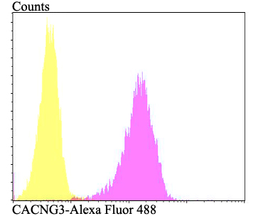
Fig4: Flow cytometric analysis of SH-SY-5Y cells with CACNG3 antibody at 1/100 dilution (fuchsia) compared with an unlabelled control (cells without incubation with primary antibody; yellow). Alexa Fluor 488-conjugated goat anti-rabbit IgG was used as the secondary antibody.Application
Fig4: Flow cytometric analysis of SH-SY-5Y cells with CACNG3 antibody at 1/100 dilution (fuchsia) compared with an unlabelled control (cells without incubation with primary antibody; yellow). Alexa Fluor 488-conjugated goat anti-rabbit IgG was used as the secondary antibody.Application
Fig4: Flow cytometric analysis of SH-SY-5Y cells with CACNG3 antibody at 1/100 dilution (fuchsia) compared with an unlabelled control (cells without incubation with primary antibody; yellow). Alexa Fluor 488-conjugated goat anti-rabbit IgG was used as the secondary antibody.Application
Fig4: Flow cytometric analysis of SH-SY-5Y cells with CACNG3 antibody at 1/100 dilution (fuchsia) compared with an unlabelled control (cells without incubation with primary antibody; yellow). Alexa Fluor 488-conjugated goat anti-rabbit IgG was used as the secondary antibody.| Product Name | CACNG3 |
|---|---|
| Antibody Type | Primary Antibodies |
| Antigen Alias | CACNG 3 antibody Cacng2 antibody CACNG3 antibody Calcium channel voltage dependent gamma subunit 3 antibody CCG3_HUMAN antibody Neuronal voltage gated calcium channel gamma 3 subunit antibody Neuronal voltage-gated calcium channel gamma-3 subunit antibody TARP gamma-3 antibody Transmembrane AMPAR regulatory protein gamma-3 antibody Voltage dependent calcium channel gamma 3 subunit antibody Voltage gated calcium channel gamma subunit antibody Voltage-dependent calcium channel gamma-3 subunit antibody |
| Product description | Regulates the trafficking to the somatodendritic compartment and gating properties of AMPA-selective glutamate receptors (AMPARs). Promotes their targeting to the cell membrane and synapses and modulates their gating properties by slowing their rates of activation, deactivation and desensitization. Does not show subunit-specific AMPA receptor regulation and regulates all AMPAR subunits. Thought to stabilize the calcium channel in an inactivated (closed) state. |
| Immunogen | Synthetic peptide within rat CACNG3 aa 200-250. |
| Clonality | Polyclonal |
|---|---|
| Isotype | IgG |
| Host Species | Rabbit |
| Tested Applications | WBICCIHCFC |
| WB:1:500 ICC:1:50-1:200 IHC:1:50-1:200 FC:1:50-1:100 |
|
| Species Reactivity | HumanRat |
| Concentration | 1mg/ml |
| Alternative Names | CACNG 3 antibody Cacng2 antibody CACNG3 antibody Calcium channel voltage dependent gamma subunit 3 antibody CCG3_HUMAN antibody Neuronal voltage gated calcium channel gamma 3 subunit antibody Neuronal voltage-gated calcium channel gamma-3 subunit antibody TARP gamma-3 antibody Transmembrane AMPAR regulatory protein gamma-3 antibody Voltage dependent calcium channel gamma 3 subunit antibody Voltage gated calcium channel gamma subunit antibody Voltage-dependent calcium channel gamma-3 subunit antibody |
|---|---|
| Molecular Weight(MW) | Predicted band size 36 kDa |
| Cellular Localization | Membrane. Displays a somatodendritic localization and is excluded from axons in neurons. |
Application
Fig4: Flow cytometric analysis of SH-SY-5Y cells with CACNG3 antibody at 1/100 dilution (fuchsia) compared with an unlabelled control (cells without incubation with primary antibody; yellow). Alexa Fluor 488-conjugated goat anti-rabbit IgG was used as the secondary antibody.
Application
Fig4: Flow cytometric analysis of SH-SY-5Y cells with CACNG3 antibody at 1/100 dilution (fuchsia) compared with an unlabelled control (cells without incubation with primary antibody; yellow). Alexa Fluor 488-conjugated goat anti-rabbit IgG was used as the secondary antibody.
Application
Fig4: Flow cytometric analysis of SH-SY-5Y cells with CACNG3 antibody at 1/100 dilution (fuchsia) compared with an unlabelled control (cells without incubation with primary antibody; yellow). Alexa Fluor 488-conjugated goat anti-rabbit IgG was used as the secondary antibody.
Application
Fig4: Flow cytometric analysis of SH-SY-5Y cells with CACNG3 antibody at 1/100 dilution (fuchsia) compared with an unlabelled control (cells without incubation with primary antibody; yellow). Alexa Fluor 488-conjugated goat anti-rabbit IgG was used as the secondary antibody.| Positive Control | SH-SY-5Y, rat brain tissue. |
|---|---|
| Application Notes | WB:1:500 ICC:1:50-1:200 IHC:1:50-1:200 FC:1:50-1:100 |
| Form | Liquid |
|---|---|
| Storage Instructions | Store at +4℃ after thawing. Aliquot store at -20℃. Avoid repeated freeze / thaw cycles. |
| Storage Buffer | 1*TBS (pH7.4), 0.5%BSA, 50%Glycerol. Preservative: 0.05% Sodium Azide. |


