Application
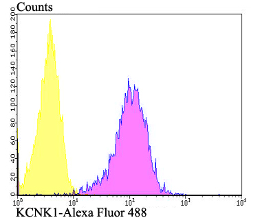
Fig7: Flow cytometric analysis of SH-SY-5Y cells with KCNK1 antibody at 1/100 dilution (fuchsia) compared with an unlabelled control (cells without incubation with primary antibody; yellow). Alexa Fluor 488-conjugated goat anti-rabbit IgG was used as the secondary antibody.Application
Fig7: Flow cytometric analysis of SH-SY-5Y cells with KCNK1 antibody at 1/100 dilution (fuchsia) compared with an unlabelled control (cells without incubation with primary antibody; yellow). Alexa Fluor 488-conjugated goat anti-rabbit IgG was used as the secondary antibody.Application
Fig7: Flow cytometric analysis of SH-SY-5Y cells with KCNK1 antibody at 1/100 dilution (fuchsia) compared with an unlabelled control (cells without incubation with primary antibody; yellow). Alexa Fluor 488-conjugated goat anti-rabbit IgG was used as the secondary antibody.Application
Fig7: Flow cytometric analysis of SH-SY-5Y cells with KCNK1 antibody at 1/100 dilution (fuchsia) compared with an unlabelled control (cells without incubation with primary antibody; yellow). Alexa Fluor 488-conjugated goat anti-rabbit IgG was used as the secondary antibody.Application
Fig7: Flow cytometric analysis of SH-SY-5Y cells with KCNK1 antibody at 1/100 dilution (fuchsia) compared with an unlabelled control (cells without incubation with primary antibody; yellow). Alexa Fluor 488-conjugated goat anti-rabbit IgG was used as the secondary antibody.Application
Fig7: Flow cytometric analysis of SH-SY-5Y cells with KCNK1 antibody at 1/100 dilution (fuchsia) compared with an unlabelled control (cells without incubation with primary antibody; yellow). Alexa Fluor 488-conjugated goat anti-rabbit IgG was used as the secondary antibody.Application
Fig7: Flow cytometric analysis of SH-SY-5Y cells with KCNK1 antibody at 1/100 dilution (fuchsia) compared with an unlabelled control (cells without incubation with primary antibody; yellow). Alexa Fluor 488-conjugated goat anti-rabbit IgG was used as the secondary antibody.| Product Name | KCNK1 |
|---|---|
| Antibody Type | Primary Antibodies |
| Antigen Alias | DPK antibody HOHO antibody HOHO1 antibody inward rectifying potassium channel protein TWIK 1 antibody Inward rectifying potassium channel protein TWIK-1 antibody Inward rectifying potassium channel protein TWIK1 antibody K2P1 antibody K2p1.1 antibody KCNK1 antibody KCNK1_HUMAN antibody KCNO1 antibody OTTHUMP00000036029 antibody Potassium channel KCNO1 antibody Potassium channel subfamily K member 1 antibody Potassium inwardly rectifying channel subfamily K member 1 antibody TWIK 1 antibody TWIK1 antibody |
| Product description | Ion channel that contributes to passive transmembrane potassium transport and to the regulation of the resting membrane potential in brain astrocytes, but also in kidney and in other tissues. Forms dimeric channels through which potassium ions pass in accordance with their electrochemical gradient. The channel is selective for K+ ions at physiological potassium concentrations and at neutral pH, but becomes permeable to Na+ at subphysiological K+ levels and upon acidification of the extracellular medium. Channel activity is modulated by activation of serotonin receptors. Heterodimeric channels containing KCNK1 and KCNK2 have much higher activity, and may represent the predominant form in astrocytes. Heterodimeric channels containing KCNK1 and KCNK3 or KCNK9 have much higher activity. Heterodimeric channels formed by KCNK1 and KCNK9 may contribute to halothane-sensitive currents. Mediates outward rectifying potassium currents in dentate gyrus granule cells and contributes to the regulation of their resting membrane potential. Contributes to the regulation of action potential firing in dentate gyrus granule cells and down-regulates their intrinsic excitability. In astrocytes, the heterodimer formed by KCNK1 and KCNK2 is required for rapid glutamate release in response to activation of G-protein coupled receptors, such as F2R and CNR1. |
| Immunogen | Synthetic peptide within human KCNK1 aa 40-100. |
| Clonality | Polyclonal |
|---|---|
| Isotype | IgG |
| Host Species | Rabbit |
| Tested Applications | WBIHCFC |
| WB:1:500 IHC:1:50-1:200 FC:1:50-1:100 |
|
| Species Reactivity | HumanMouseRat |
| Concentration | 1mg/ml |
| Alternative Names | DPK antibody HOHO antibody HOHO1 antibody inward rectifying potassium channel protein TWIK 1 antibody Inward rectifying potassium channel protein TWIK-1 antibody Inward rectifying potassium channel protein TWIK1 antibody K2P1 antibody K2p1.1 antibody KCNK1 antibody KCNK1_HUMAN antibody KCNO1 antibody OTTHUMP00000036029 antibody Potassium channel KCNO1 antibody Potassium channel subfamily K member 1 antibody Potassium inwardly rectifying channel subfamily K member 1 antibody TWIK 1 antibody TWIK1 antibody |
|---|---|
| Molecular Weight(MW) | 38 kDa |
| Cellular Localization | Cell junction. Cell membrane. Cell projection. Cytoplasmic vesicle. Endosome. Membrane. Synapse. |
Application
Fig7: Flow cytometric analysis of SH-SY-5Y cells with KCNK1 antibody at 1/100 dilution (fuchsia) compared with an unlabelled control (cells without incubation with primary antibody; yellow). Alexa Fluor 488-conjugated goat anti-rabbit IgG was used as the secondary antibody.
Application
Fig7: Flow cytometric analysis of SH-SY-5Y cells with KCNK1 antibody at 1/100 dilution (fuchsia) compared with an unlabelled control (cells without incubation with primary antibody; yellow). Alexa Fluor 488-conjugated goat anti-rabbit IgG was used as the secondary antibody.
Application
Fig7: Flow cytometric analysis of SH-SY-5Y cells with KCNK1 antibody at 1/100 dilution (fuchsia) compared with an unlabelled control (cells without incubation with primary antibody; yellow). Alexa Fluor 488-conjugated goat anti-rabbit IgG was used as the secondary antibody.
Application
Fig7: Flow cytometric analysis of SH-SY-5Y cells with KCNK1 antibody at 1/100 dilution (fuchsia) compared with an unlabelled control (cells without incubation with primary antibody; yellow). Alexa Fluor 488-conjugated goat anti-rabbit IgG was used as the secondary antibody.
Application
Fig7: Flow cytometric analysis of SH-SY-5Y cells with KCNK1 antibody at 1/100 dilution (fuchsia) compared with an unlabelled control (cells without incubation with primary antibody; yellow). Alexa Fluor 488-conjugated goat anti-rabbit IgG was used as the secondary antibody.
Application
Fig7: Flow cytometric analysis of SH-SY-5Y cells with KCNK1 antibody at 1/100 dilution (fuchsia) compared with an unlabelled control (cells without incubation with primary antibody; yellow). Alexa Fluor 488-conjugated goat anti-rabbit IgG was used as the secondary antibody.
Application
Fig7: Flow cytometric analysis of SH-SY-5Y cells with KCNK1 antibody at 1/100 dilution (fuchsia) compared with an unlabelled control (cells without incubation with primary antibody; yellow). Alexa Fluor 488-conjugated goat anti-rabbit IgG was used as the secondary antibody.| Positive Control | Human placenta tissue, human liver tissue, rat cerebellum tissue, human kidney tissue, mouse brain tissue, SH-SY-5Y. |
|---|---|
| Application Notes | WB:1:500 IHC:1:50-1:200 FC:1:50-1:100 |
| Form | Liquid |
|---|---|
| Storage Instructions | Store at +4℃ after thawing. Aliquot store at -20℃. Avoid repeated freeze / thaw cycles. |
| Storage Buffer | 1*TBS (pH7.4), 0.5%BSA, 50%Glycerol. Preservative: 0.05% Sodium Azide. |


