Application
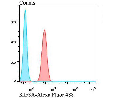
Fig4: Flow cytometric analysis of A549 cells with KIF3A antibody at 1/100 dilution (red) compared with an unlabelled control (cells without incubation with primary antibody; blue). Alexa Fluor 488-conjugated goat anti-rabbit IgG was used as the secondary antibody.Application
Fig4: Flow cytometric analysis of A549 cells with KIF3A antibody at 1/100 dilution (red) compared with an unlabelled control (cells without incubation with primary antibody; blue). Alexa Fluor 488-conjugated goat anti-rabbit IgG was used as the secondary antibody.Application
Fig4: Flow cytometric analysis of A549 cells with KIF3A antibody at 1/100 dilution (red) compared with an unlabelled control (cells without incubation with primary antibody; blue). Alexa Fluor 488-conjugated goat anti-rabbit IgG was used as the secondary antibody.Application
Fig4: Flow cytometric analysis of A549 cells with KIF3A antibody at 1/100 dilution (red) compared with an unlabelled control (cells without incubation with primary antibody; blue). Alexa Fluor 488-conjugated goat anti-rabbit IgG was used as the secondary antibody.| Product Name | KIF3A |
|---|---|
| Antibody Type | Primary Antibodies |
| Antigen Alias | KIF 3 antibody KIF 3A antibody KIF3 antibody Kif3a antibody KIF3A_HUMAN antibody Kifl antibody Kinesin family member 3A antibody Kinesin family protein 3 antibody Kinesin family protein 3A antibody Kinesin like protein KIF 3A antibody Kinesin like protein KIF3A antibody Kinesin-like protein KIF3A antibody Kns3 antibody Microtubule plus end directed kinesin motor 3A antibody Microtubule plus end-directed kinesin motor 3A antibody |
| Product description | Microtubule-based anterograde translocator for membranous organelles. Plus end-directed microtubule sliding activity in vitro. Plays a role in primary cilia formation. Plays a role in centriole cohesion and subdistal appendage organization and function. Regulates the formation of the subdistal appendage via recruitement of DCTN1 to the centriole. Also required for ciliary basal feet formation and microtubule anchoring to mother centriole. |
| Immunogen | Recombinant protein within human KIF3A aa 100-360. |
| Clonality | Polyclonal |
|---|---|
| Isotype | IgG |
| Host Species | Rabbit |
| Tested Applications | WBIHCFC |
| WB:1:500-1:1,000 IHC:1:50-1:100 FC:1:50-1:100 |
|
| Species Reactivity | HumanMouseRat |
| Concentration | 1mg/ml |
| Alternative Names | KIF 3 antibody KIF 3A antibody KIF3 antibody Kif3a antibody KIF3A_HUMAN antibody Kifl antibody Kinesin family member 3A antibody Kinesin family protein 3 antibody Kinesin family protein 3A antibody Kinesin like protein KIF 3A antibody Kinesin like protein KIF3A antibody Kinesin-like protein KIF3A antibody Kns3 antibody Microtubule plus end directed kinesin motor 3A antibody Microtubule plus end-directed kinesin motor 3A antibody |
|---|---|
| Molecular Weight(MW) | 80 kDa |
| Cellular Localization | Cytoskeleton. |
Application
Fig4: Flow cytometric analysis of A549 cells with KIF3A antibody at 1/100 dilution (red) compared with an unlabelled control (cells without incubation with primary antibody; blue). Alexa Fluor 488-conjugated goat anti-rabbit IgG was used as the secondary antibody.
Application
Fig4: Flow cytometric analysis of A549 cells with KIF3A antibody at 1/100 dilution (red) compared with an unlabelled control (cells without incubation with primary antibody; blue). Alexa Fluor 488-conjugated goat anti-rabbit IgG was used as the secondary antibody.
Application
Fig4: Flow cytometric analysis of A549 cells with KIF3A antibody at 1/100 dilution (red) compared with an unlabelled control (cells without incubation with primary antibody; blue). Alexa Fluor 488-conjugated goat anti-rabbit IgG was used as the secondary antibody.
Application
Fig4: Flow cytometric analysis of A549 cells with KIF3A antibody at 1/100 dilution (red) compared with an unlabelled control (cells without incubation with primary antibody; blue). Alexa Fluor 488-conjugated goat anti-rabbit IgG was used as the secondary antibody.| Positive Control | Rat testis tissue, Daudi, rat cerebellum tissue, mouse testis tissue, A549. |
|---|---|
| Application Notes | WB:1:500-1:1,000 IHC:1:50-1:100 FC:1:50-1:100 |
| Form | Liquid |
|---|---|
| Storage Instructions | Store at +4℃ after thawing. Aliquot store at -20℃. Avoid repeated freeze / thaw cycles. |
| Storage Buffer | 1*TBS (pH7.4), 0.5%BSA, 50%Glycerol. Preservative: 0.05% Sodium Azide. |


