Application
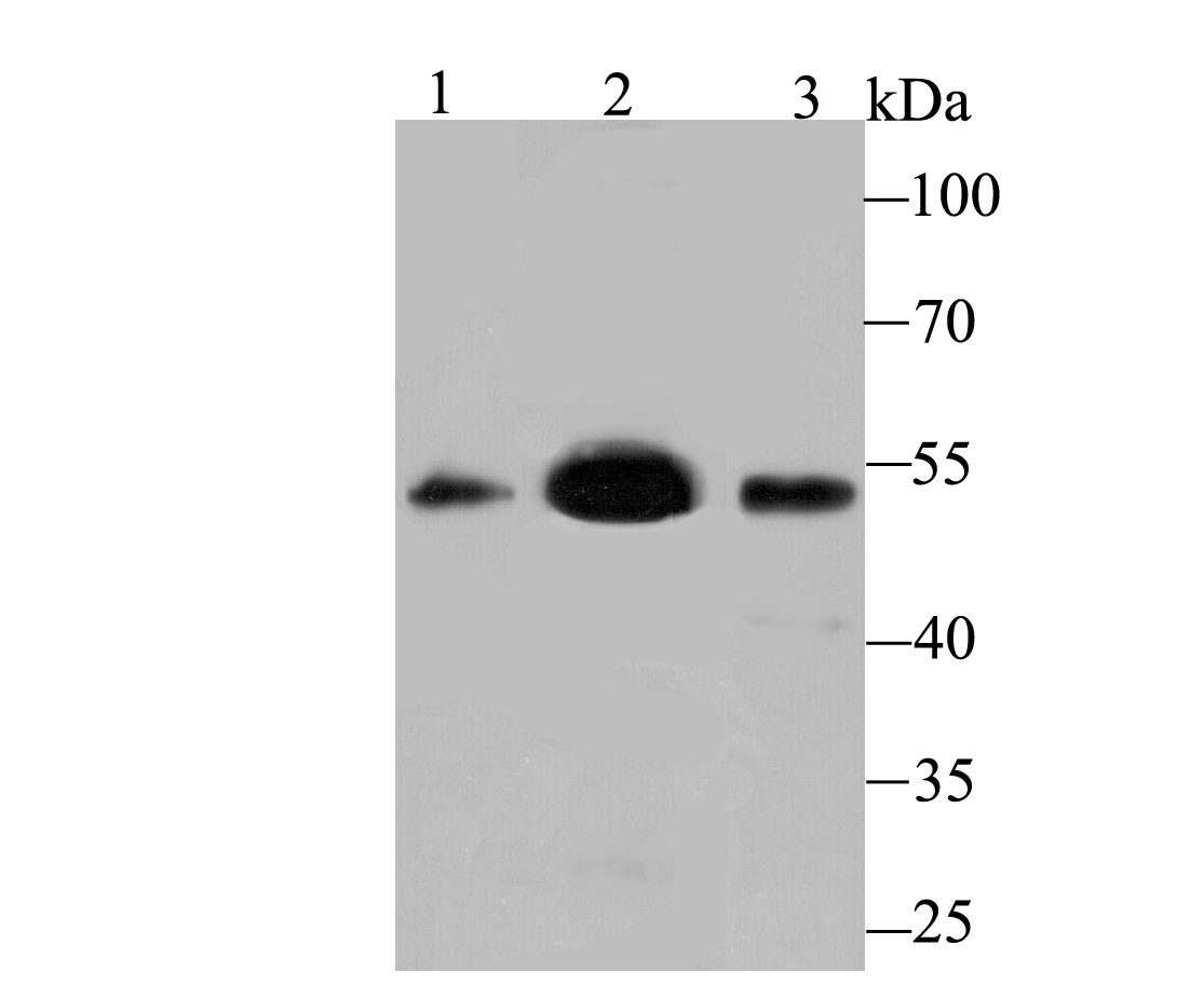
Fig1: Western blot analysis of MEIS2 on different lysates using anti-MEIS2 antibody at 1/500 dilution. Positive control: Lane 1: Mouse testis Lane 2: SH-SY-5Y Lane 3: Mouse lungApplication
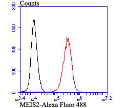
Fig2: Flow cytometric analysis of K562 cells with MEIS2 antibody at 1/100 dilution (red) compared with an unlabelled control (cells without incubation with primary antibody; black). Alexa Fluor 488-conjugated goat anti-rabbit IgG was used as the secondary antibody.| Product Name | MEIS2 [JB40-38] |
|---|---|
| Antibody Type | Primary Antibodies |
| Antigen Alias | Homeobox protein Meis2 antibody HsT18361 antibody Meis (mouse) homolog 2 antibody Meis homeobox 2 antibody Meis homolog 2 antibody Meis1 myeloid ecotropic viral integration site 1 homolog 2 (mouse) antibody Meis1 myeloid ecotropic viral integration site 1 homolog 2 antibody Meis1 related gene 1 antibody Meis1 related protein 1 antibody Meis1-related protein 1 antibody MEIS2 antibody MEIS2_HUMAN antibody MGC2820 antibody MRG1 antibody TALE homeobox protein Meis2 antibody |
| Product description | Profilins regulate Actin polymerization by binding to and sequestering the Actin monomer. Profilins act as a nucleotide exchange factor that charges Actin with ATP after binding the Actin monomer through a 1:1 stoichiometric relationship. Human Profilin-1 and Profilin-2 are encoded by two separate genes mapping to chromosomes 17p13.2 and 3q25.1, respectively. Both Profilin-1 and Profilin-2 are abundantly expressed in kidney. Profilin-1 is highly expressed in lung, liver, placenta and kidney while Profilin-2 is highly expressed in brain and skeletal muscle. In axonal and dendritic processes of mouse brain, Profilins co-localize with dynamin I and synapsin. Profilin may play a role in mediating cell adhesion. The overexpression of Profilin in endothelial cells results in increased adhesion to Fibronectin. In food allergy, plant Profilin is considered a pan-allergen. Case studies indicate individuals with allergies to various foods including celery, carrots, zucchini and peanuts are actually sensitive to the Profilin proteins in these foods. |
| Immunogen | Recombinant protein |
| Clonality | Monoclonal |
|---|---|
| Isotype | IgG |
| Host Species | Recombinant rabbit |
| Tested Applications | WBFC |
| WB:1:500-1:1,000 FC:1:50-1:100 |
|
| Species Reactivity | HumanMouse |
| Concentration | 1mg/ml |
| Alternative Names | Homeobox protein Meis2 antibody HsT18361 antibody Meis (mouse) homolog 2 antibody Meis homeobox 2 antibody Meis homolog 2 antibody Meis1 myeloid ecotropic viral integration site 1 homolog 2 (mouse) antibody Meis1 myeloid ecotropic viral integration site 1 homolog 2 antibody Meis1 related gene 1 antibody Meis1 related protein 1 antibody Meis1-related protein 1 antibody MEIS2 antibody MEIS2_HUMAN antibody MGC2820 antibody MRG1 antibody TALE homeobox protein Meis2 antibody |
|---|---|
| Molecular Weight(MW) | 51 kDa |
| Cellular Localization | Nucleus. Cytoplasm. |
Application
Fig1: Western blot analysis of MEIS2 on different lysates using anti-MEIS2 antibody at 1/500 dilution. Positive control: Lane 1: Mouse testis Lane 2: SH-SY-5Y Lane 3: Mouse lung
Application
Fig2: Flow cytometric analysis of K562 cells with MEIS2 antibody at 1/100 dilution (red) compared with an unlabelled control (cells without incubation with primary antibody; black). Alexa Fluor 488-conjugated goat anti-rabbit IgG was used as the secondary antibody.| Positive Control | Mouse testis tissue lysate, SH-SY-5Y, human kidney tissue lysate, mouse lung tissue lysate, K562. |
|---|---|
| Application Notes | WB:1:500-1:1,000 FC:1:50-1:100 |
| Form | Liquid |
|---|---|
| Storage Instructions | Store at +4℃ after thawing. Aliquot store at -20℃ or -80℃. Avoid repeated freeze / thaw cycles. |
| Storage Buffer | 1*TBS (pH7.4), 1%BSA, 40%Glycerol. Preservative: 0.05% Sodium Azide. |


