Application
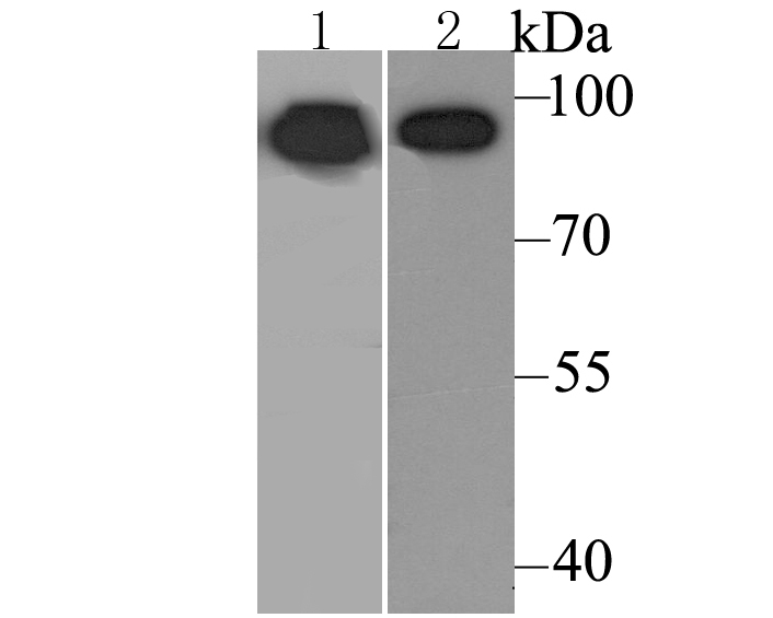
Fig1: Western blot analysis of Calnexin on mouse lung tissue (1) and Daudi cell (2) lysate using anti-Calnexin antibody at 1/500 dilution.Application
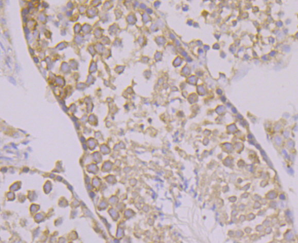
Fig2: Immunohistochemical analysis of paraffin-embedded mouse testis tissue using anti-Calnexin antibody. Counter stained with hematoxylin.Application
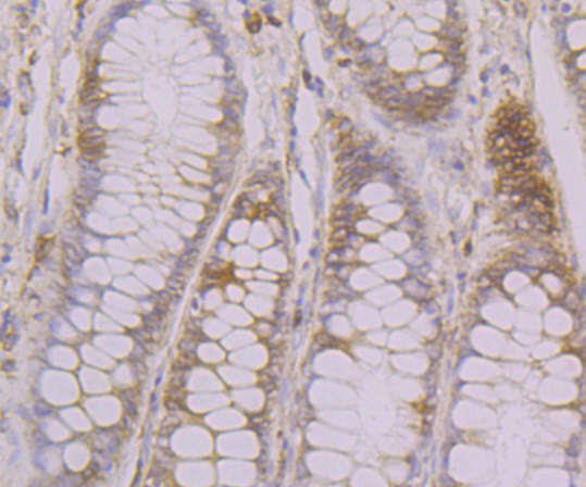
Fig3: Immunohistochemical analysis of paraffin-embedded human colon tissue using anti-Calnexin antibody. Counter stained with hematoxylin.Application
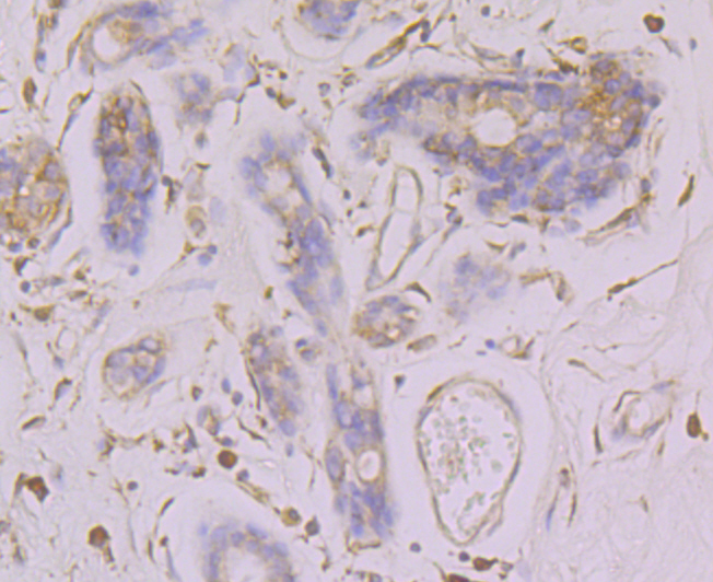
Fig4: Immunohistochemical analysis of paraffin-embedded human breast cancer tissue using anti-Calnexin antibody. Counter stained with hematoxylin.Application
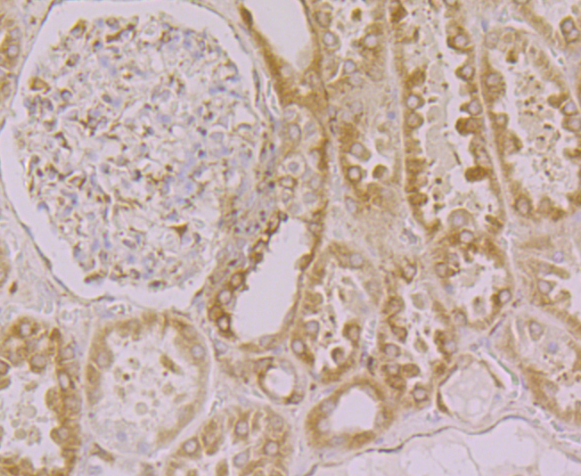
Fig5: Immunohistochemical analysis of paraffin-embedded human kidney tissue using anti-Calnexin antibody. Counter stained with hematoxylin.Application
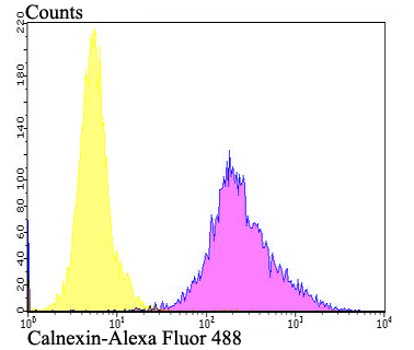
Fig6: Flow cytometric analysis of SKOV-3 cells with Calnexin antibody at 1/100 dilution (fuchsia) compared with an unlabelled control (cells without incubation with primary antibody; yellow). Alexa Fluor 488-conjugated goat anti-rabbit IgG was used as the secondary antibody.| Product Name | Calnexin |
|---|---|
| Antibody Type | Primary Antibodies |
| Antigen Alias | Calnexin antibody CALX_HUMAN antibody CANX antibody CNX antibody FLJ26570 antibody Histocompatibility complex class I antigen binding protein p88 antibody IP90 antibody Major histocompatibility complex class I antigen-binding protein p88 antibody p90 antibody |
| Product description | Calnexin and Calregulin (also called calreticulin) are calcium-binding proteins that are localized to the endoplasmic reticulum, Calnexin to the membrane and Calregulin to the lumen. Calnexin is a type I membrane protein that interacts with newly synthesized glycoproteins in the endoplasmic reticulum. It may play a role in assisting with protein assembly and in retaining unassembled protein subunits in the endoplasmic reticulum. Calregulin has both low- and high-affinity calcium-binding sites. Neither Calnexin nor Calregulin contains the calcium-binding “E-F hand” motif found in calmodulins. Calnexin and Calregulin are important for the maturation of glycoproteins in the endoplasmic reticulum and appear to bind many of the same proteins. |
| Immunogen | Synthetic peptide within C-terminal residues of rat Calnexin. |
| Clonality | Polyclonal |
|---|---|
| Isotype | IgG |
| Host Species | Rabbit |
| Tested Applications | WBIHCFC |
| WB:1:500-1:2,000 IHC:1:50-1:200 FC:1:50-1:100 |
|
| Species Reactivity | HumanMouse |
| Concentration | 1mg/ml |
| Alternative Names | Calnexin antibody CALX_HUMAN antibody CANX antibody CNX antibody FLJ26570 antibody Histocompatibility complex class I antigen binding protein p88 antibody IP90 antibody Major histocompatibility complex class I antigen-binding protein p88 antibody p90 antibody |
|---|---|
| Molecular Weight(MW) | 90 kDa |
| Cellular Localization | Endoplasmic reticulum. |
Application
Fig1: Western blot analysis of Calnexin on mouse lung tissue (1) and Daudi cell (2) lysate using anti-Calnexin antibody at 1/500 dilution.
Application
Fig2: Immunohistochemical analysis of paraffin-embedded mouse testis tissue using anti-Calnexin antibody. Counter stained with hematoxylin.
Application
Fig3: Immunohistochemical analysis of paraffin-embedded human colon tissue using anti-Calnexin antibody. Counter stained with hematoxylin.
Application
Fig4: Immunohistochemical analysis of paraffin-embedded human breast cancer tissue using anti-Calnexin antibody. Counter stained with hematoxylin.
Application
Fig5: Immunohistochemical analysis of paraffin-embedded human kidney tissue using anti-Calnexin antibody. Counter stained with hematoxylin.
Application
Fig6: Flow cytometric analysis of SKOV-3 cells with Calnexin antibody at 1/100 dilution (fuchsia) compared with an unlabelled control (cells without incubation with primary antibody; yellow). Alexa Fluor 488-conjugated goat anti-rabbit IgG was used as the secondary antibody.| Positive Control | Daudi, mouse lung tissue, human colon tissue, human breast cancer tissue, human kidney tissue, mouse testis tissue, SKOV-3. |
|---|---|
| Application Notes | WB:1:500-1:2,000 IHC:1:50-1:200 FC:1:50-1:100 |
| Form | Liquid |
|---|---|
| Storage Instructions | Store at +4℃ after thawing. Aliquot store at -20℃. Avoid repeated freeze / thaw cycles. |
| Storage Buffer | 1*TBS (pH7.4), 0.5%BSA, 50%Glycerol. Preservative: 0.05% Sodium Azide. |


