Application
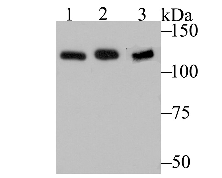
Fig1: Western blot analysis of HnRNP U on different cell lysates using anti-HnRNP U at 1/500 dilution. Positive control: Lane 1: NIH-3T3 Lane 2: SH-SY-5Y Lane 3: K562Application
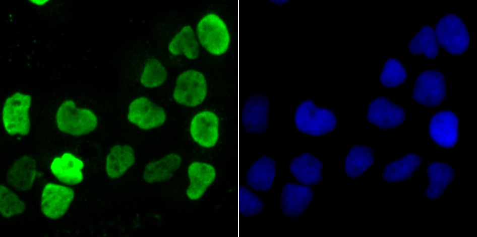
Fig2: ICC staining HnRNP U in A431 cells (green). The nuclear counter stain is DAPI (blue). Cells were fixed in paraformaldehyde, permeabilised with 0.25% Triton X100/PBS.Application
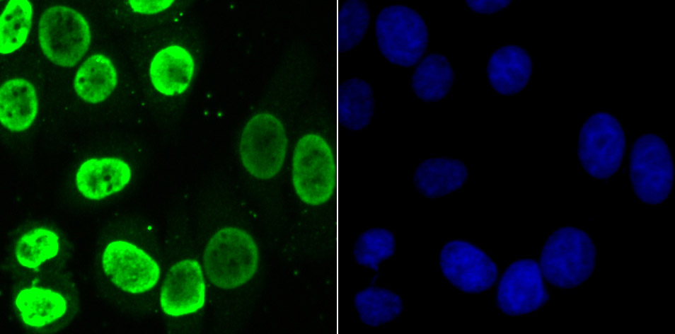
Fig3: ICC staining HnRNP U in HepG2 cells (green). The nuclear counter stain is DAPI (blue). Cells were fixed in paraformaldehyde, permeabilised with 0.25% Triton X100/PBS.Application
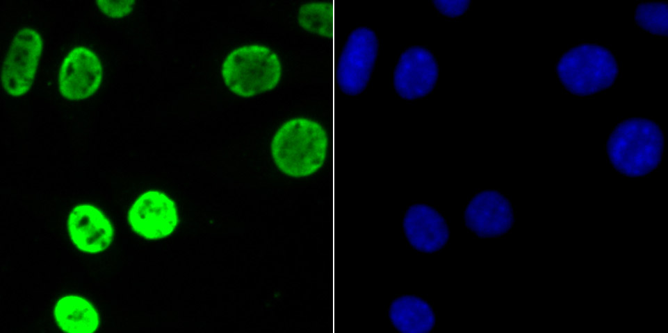
Fig4: ICC staining HnRNP U in SHSY-5Y cells (green). The nuclear counter stain is DAPI (blue). Cells were fixed in paraformaldehyde, permeabilised with 0.25% Triton X100/PBS.Application
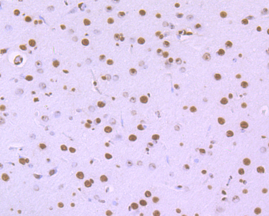
Fig5: Immunohistochemical analysis of paraffin-embedded rat brain tissue using anti-HnRNP U antibody. Counter stained with hematoxylin.Application
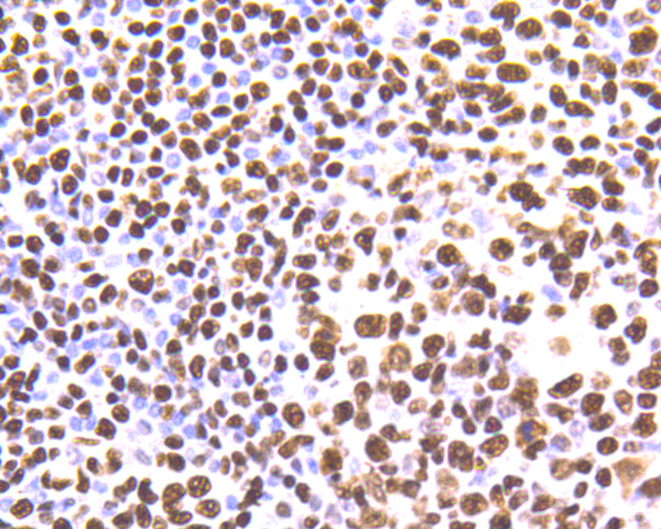
Fig6: Immunohistochemical analysis of paraffin-embedded human tonsil tissue using anti-HnRNP U antibody. Counter stained with hematoxylin.Application
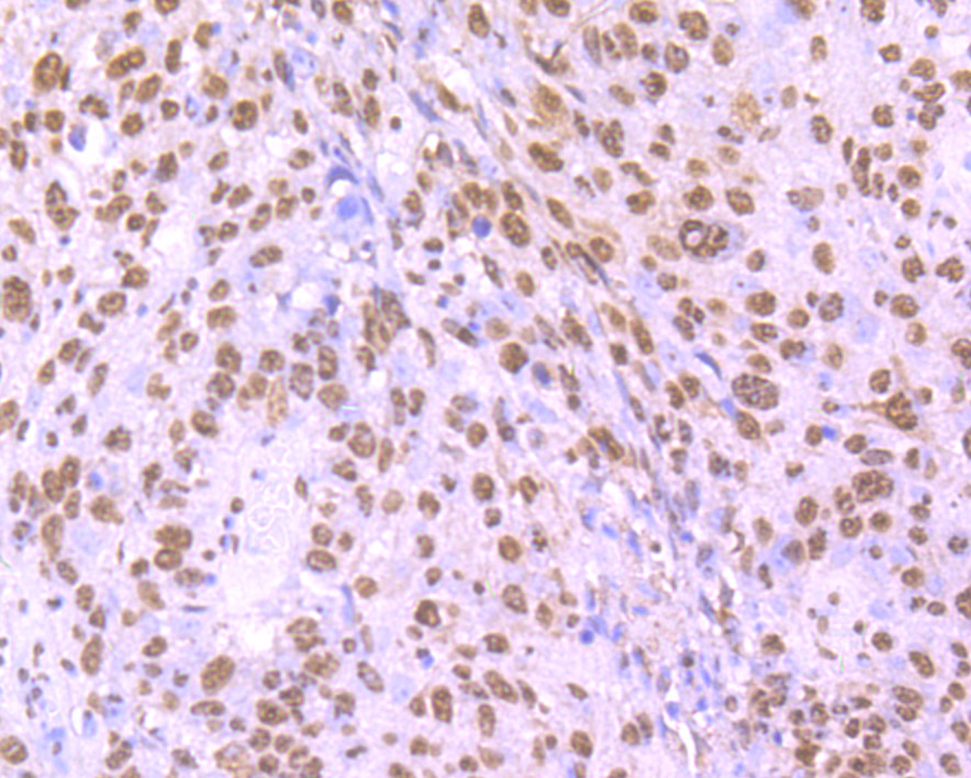
Fig7: Immunohistochemical analysis of paraffin-embedded human lung cancer tissue using anti-HnRNP U antibody. Counter stained with hematoxylin.Application
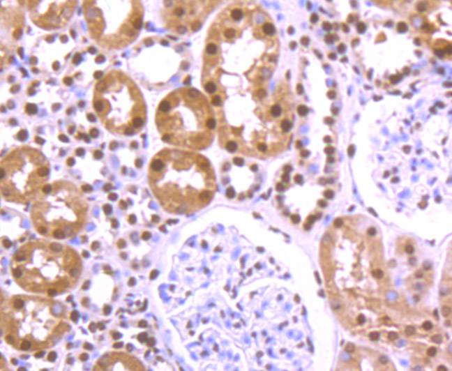
Fig8: Immunohistochemical analysis of paraffin-embedded human kidney tissue using anti-HnRNP U antibody. Counter stained with hematoxylin.Application
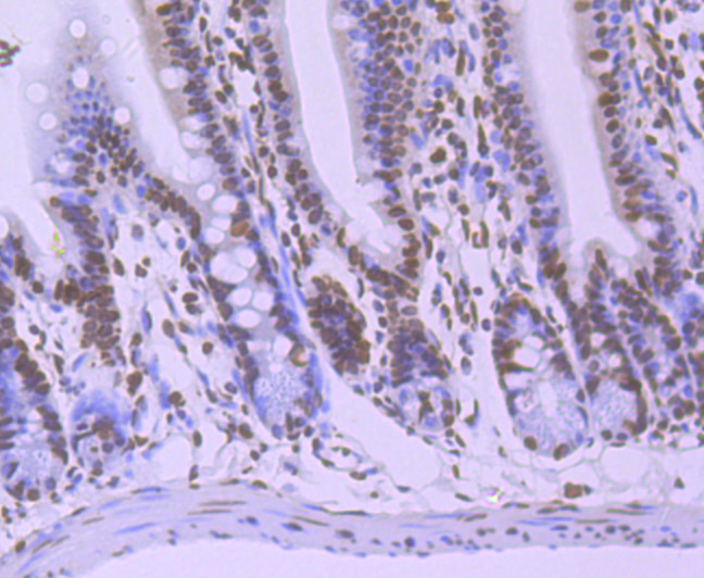
Fig9: Immunohistochemical analysis of paraffin-embedded mouse colon tissue using anti-HnRNP U antibody. Counter stained with hematoxylin.| Product Name | hnRNP U [JB23-43] |
|---|---|
| Antibody Type | Primary Antibodies |
| Antigen Alias | Heterogeneous nuclear ribonucleoprotein U antibody hnRNP U antibody hnRNP U protein antibody HNRNPU antibody hnRNPU protein antibody HNRPU antibody HNRPU_HUMAN antibody p120 antibody p120 nuclear protein antibody pp120 antibody SAF A antibody SAF-A antibody SAFA antibody Scaffold attachment factor A antibody U21.1 antibody |
| Product description | DNA- and RNA-binding protein involved in several cellular processes such as nuclear chromatin organization, telomere-length regulation, transcription, mRNA alternative splicing and stability, Xist-mediated transcriptional silencing and mitotic cell progression. Plays a role in the regulation of interphase large-scale gene-rich chromatin organization through chromatin-associated RNAs (caRNAs) in a transcription-dependent manner, and thereby maintains genomic stability. Plays a role as a RNA polymerase II (Pol II) holoenzyme transcription regulator. Promotes transcription initiation by direct association with the core-TFIIH basal transcription factor complex for the assembly of a functional pre-initiation complex with Pol II in a actin-dependent manner. Blocks Pol II transcription elongation activity by inhibiting the C-terminal domain (CTD) phosphorylation of Pol II and dissociates from Pol II pre-initiation complex prior to productive transcription elongation. Positively regulates CBX5-induced transcriptional gene silencing and retention of CBX5 in the nucleus. |
| Immunogen | Recombinant protein |
| Clonality | Monoclonal |
|---|---|
| Isotype | IgG |
| Host Species | Recombinant rabbit |
| Tested Applications | WBICCIFIHC |
| WB:1:500-1:1,000 ICC:1:100-1:500 IHC:1:50-1:200 |
|
| Species Reactivity | HumanMouseRat |
| Concentration | 1mg/ml |
| Alternative Names | Heterogeneous nuclear ribonucleoprotein U antibody hnRNP U antibody hnRNP U protein antibody HNRNPU antibody hnRNPU protein antibody HNRPU antibody HNRPU_HUMAN antibody p120 antibody p120 nuclear protein antibody pp120 antibody SAF A antibody SAF-A antibody SAFA antibody Scaffold attachment factor A antibody U21.1 antibody |
|---|---|
| Molecular Weight(MW) | 120 kDa |
| Cellular Localization | Nucleus. Cytoskeleton. |
Application
Fig1: Western blot analysis of HnRNP U on different cell lysates using anti-HnRNP U at 1/500 dilution. Positive control: Lane 1: NIH-3T3 Lane 2: SH-SY-5Y Lane 3: K562
Application
Fig2: ICC staining HnRNP U in A431 cells (green). The nuclear counter stain is DAPI (blue). Cells were fixed in paraformaldehyde, permeabilised with 0.25% Triton X100/PBS.
Application
Fig3: ICC staining HnRNP U in HepG2 cells (green). The nuclear counter stain is DAPI (blue). Cells were fixed in paraformaldehyde, permeabilised with 0.25% Triton X100/PBS.
Application
Fig4: ICC staining HnRNP U in SHSY-5Y cells (green). The nuclear counter stain is DAPI (blue). Cells were fixed in paraformaldehyde, permeabilised with 0.25% Triton X100/PBS.
Application
Fig5: Immunohistochemical analysis of paraffin-embedded rat brain tissue using anti-HnRNP U antibody. Counter stained with hematoxylin.
Application
Fig6: Immunohistochemical analysis of paraffin-embedded human tonsil tissue using anti-HnRNP U antibody. Counter stained with hematoxylin.
Application
Fig7: Immunohistochemical analysis of paraffin-embedded human lung cancer tissue using anti-HnRNP U antibody. Counter stained with hematoxylin.
Application
Fig8: Immunohistochemical analysis of paraffin-embedded human kidney tissue using anti-HnRNP U antibody. Counter stained with hematoxylin.
Application
Fig9: Immunohistochemical analysis of paraffin-embedded mouse colon tissue using anti-HnRNP U antibody. Counter stained with hematoxylin.| Positive Control | A431, HepG2, SH-SY-5Y, human tonsil tissue, human kidney tissue, human lung cancer tissue, mouse colon tissue, rat brain tissue, NIH-3T3. |
|---|---|
| Application Notes | WB:1:500-1:1,000 ICC:1:100-1:500 IHC:1:50-1:200 |
| Form | Liquid |
|---|---|
| Storage Instructions | Store at +4℃ after thawing. Aliquot store at -20℃ or -80℃. Avoid repeated freeze / thaw cycles. |
| Storage Buffer | 1*TBS (pH7.4), 1%BSA, 40%Glycerol. Preservative: 0.05% Sodium Azide. |


