Application
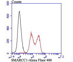
Fig4: Flow cytometric analysis of K562 cells with SMARCC1 antibody at 1/50 dilution (red) compared with an unlabelled control (cells without incubation with primary antibody; black). Alexa Fluor 488-conjugated goat anti-rabbit IgG was used as the secondary antibody.Application
Fig4: Flow cytometric analysis of K562 cells with SMARCC1 antibody at 1/50 dilution (red) compared with an unlabelled control (cells without incubation with primary antibody; black). Alexa Fluor 488-conjugated goat anti-rabbit IgG was used as the secondary antibody.Application
Fig4: Flow cytometric analysis of K562 cells with SMARCC1 antibody at 1/50 dilution (red) compared with an unlabelled control (cells without incubation with primary antibody; black). Alexa Fluor 488-conjugated goat anti-rabbit IgG was used as the secondary antibody.Application
Fig4: Flow cytometric analysis of K562 cells with SMARCC1 antibody at 1/50 dilution (red) compared with an unlabelled control (cells without incubation with primary antibody; black). Alexa Fluor 488-conjugated goat anti-rabbit IgG was used as the secondary antibody.| Product Name | SMARCC1 [JB43-43] |
|---|---|
| Antibody Type | Primary Antibodies |
| Antigen Alias | AI115498 antibody BAF 155 antibody BAF155 antibody BRG 1 associated factor 155 antibody BRG1 associated factor 155 antibody BRG1-associated factor 155 antibody Chromatin remodeling complex BAF155 subunit antibody CRACC 1 antibody CRACC1 antibody Mammalian chromatin remodeling complex BRG 1 associated factor 155 antibody Mammalian chromatin remodeling complex BRG1 associated factor 155 antibody Rsc 8 antibody Rsc8 antibody SMARC C1 antibody SMARCC 1 antibody SMARCC1 antibody SMRC1_HUMAN antibody SRG 3 antibody SRG3 antibody SWI 3 antibody SWI/SNF complex 155 kDa subunit antibody SWI/SNF complex subunit SMARCC1 antibody SWI/SNF related matrix associated actin dependent regulator of chromatin c1 antibody SWI/SNF related matrix associated actin dependent regulator of chromatin subfamily c member 1 antibody SWI/SNF-related matrix-associated actin-dependent regulator of chromatin subfamily C member 1 antibody SWI3 antibody |
| Product description | Involved in transcriptional activation and repression of select genes by chromatin remodeling (alteration of DNA-nucleosome topology). Component of SWI/SNF chromatin remodeling complexes that carry out key enzymatic activities, changing chromatin structure by altering DNA-histone contacts within a nucleosome in an ATP-dependent manner. May stimulate the ATPase activity of the catalytic subunit of the complex. Belongs to the neural progenitors-specific chromatin remodeling complex (npBAF complex) and the neuron-specific chromatin remodeling complex (nBAF complex). During neural development a switch from a stem/progenitor to a postmitotic chromatin remodeling mechanism occurs as neurons exit the cell cycle and become committed to their adult state. The transition from proliferating neural stem/progenitor cells to postmitotic neurons requires a switch in subunit composition of the npBAF and nBAF complexes. |
| Immunogen | Recombinant protein |
| Clonality | Monoclonal |
|---|---|
| Isotype | IgG |
| Host Species | Recombinant rabbit |
| Tested Applications | WBICCIFFCIP |
| WB:1:500-1:1,000 ICC:1:50-1:200 FC:1:50-1:100 IP:1:10-1:50 |
|
| Species Reactivity | HumanRat |
| Concentration | 1mg/ml |
| Alternative Names | AI115498 antibody BAF 155 antibody BAF155 antibody BRG 1 associated factor 155 antibody BRG1 associated factor 155 antibody BRG1-associated factor 155 antibody Chromatin remodeling complex BAF155 subunit antibody CRACC 1 antibody CRACC1 antibody Mammalian chromatin remodeling complex BRG 1 associated factor 155 antibody Mammalian chromatin remodeling complex BRG1 associated factor 155 antibody Rsc 8 antibody Rsc8 antibody SMARC C1 antibody SMARCC 1 antibody SMARCC1 antibody SMRC1_HUMAN antibody SRG 3 antibody SRG3 antibody SWI 3 antibody SWI/SNF complex 155 kDa subunit antibody SWI/SNF complex subunit SMARCC1 antibody SWI/SNF related matrix associated actin dependent regulator of chromatin c1 antibody SWI/SNF related matrix associated actin dependent regulator of chromatin subfamily c member 1 antibody SWI/SNF-related matrix-associated actin-dependent regulator of chromatin subfamily C member 1 antibody SWI3 antibody |
|---|---|
| Molecular Weight(MW) | 123 kDa (Predicted band size) 150 kDa (Observed band size) |
| Cellular Localization | Nucleus. Cytoplasm. |
Application
Fig4: Flow cytometric analysis of K562 cells with SMARCC1 antibody at 1/50 dilution (red) compared with an unlabelled control (cells without incubation with primary antibody; black). Alexa Fluor 488-conjugated goat anti-rabbit IgG was used as the secondary antibody.
Application
Fig4: Flow cytometric analysis of K562 cells with SMARCC1 antibody at 1/50 dilution (red) compared with an unlabelled control (cells without incubation with primary antibody; black). Alexa Fluor 488-conjugated goat anti-rabbit IgG was used as the secondary antibody.
Application
Fig4: Flow cytometric analysis of K562 cells with SMARCC1 antibody at 1/50 dilution (red) compared with an unlabelled control (cells without incubation with primary antibody; black). Alexa Fluor 488-conjugated goat anti-rabbit IgG was used as the secondary antibody.
Application
Fig4: Flow cytometric analysis of K562 cells with SMARCC1 antibody at 1/50 dilution (red) compared with an unlabelled control (cells without incubation with primary antibody; black). Alexa Fluor 488-conjugated goat anti-rabbit IgG was used as the secondary antibody.| Positive Control | Mouse thymus tissue, HL-60, 293T, mouse testis tissue, K562. |
|---|---|
| Application Notes | WB:1:500-1:1,000 ICC:1:50-1:200 FC:1:50-1:100 IP:1:10-1:50 |
| Form | Liquid |
|---|---|
| Storage Instructions | Store at +4℃ after thawing. Aliquot store at -20℃ or -80℃. Avoid repeated freeze / thaw cycles. |
| Storage Buffer | 1*TBS (pH7.4), 1%BSA, 40%Glycerol. Preservative: 0.05% Sodium Azide. |


