Application
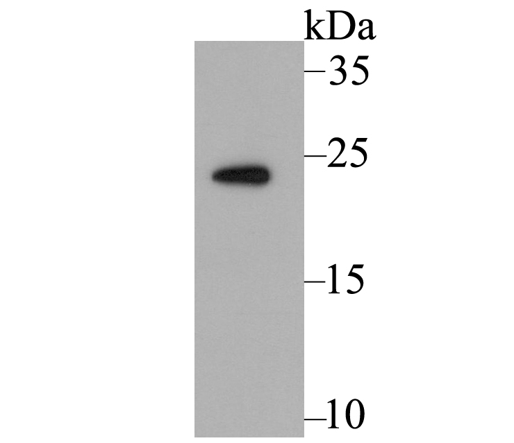
Fig1: Western blot analysis of Methyltransferase-like 26 on mouse kidney tissue lysate using anti-Methyltransferase-like 26 antibody at 1/500 dilution.Application
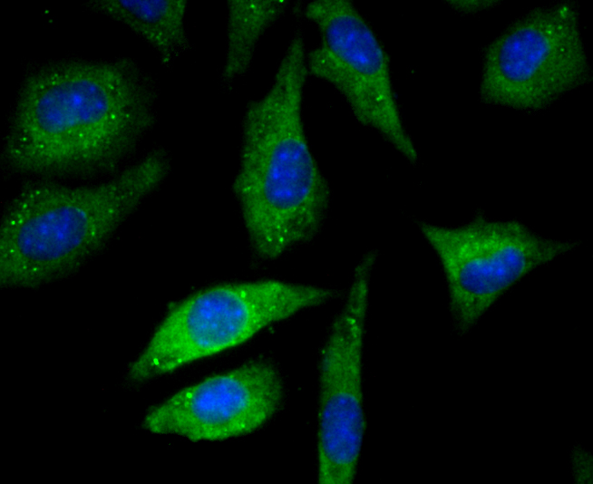
Fig2: ICC staining Methyltransferase-like 26 in SH-SY-5Y cells (green). The nuclear counter stain is DAPI (blue). Cells were fixed in paraformaldehyde, permeabilised with 0.25% Triton X100/PBS.Application
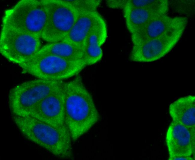
Fig3: ICC staining Methyltransferase-like 26 in MCF-7 cells (green). The nuclear counter stain is DAPI (blue). Cells were fixed in paraformaldehyde, permeabilised with 0.25% Triton X100/PBS.Application
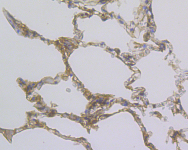
Fig4: Immunohistochemical analysis of paraffin-embedded human lung tissue using anti-Methyltransferase-like 26 antibody. Counter stained with hematoxylin.Application
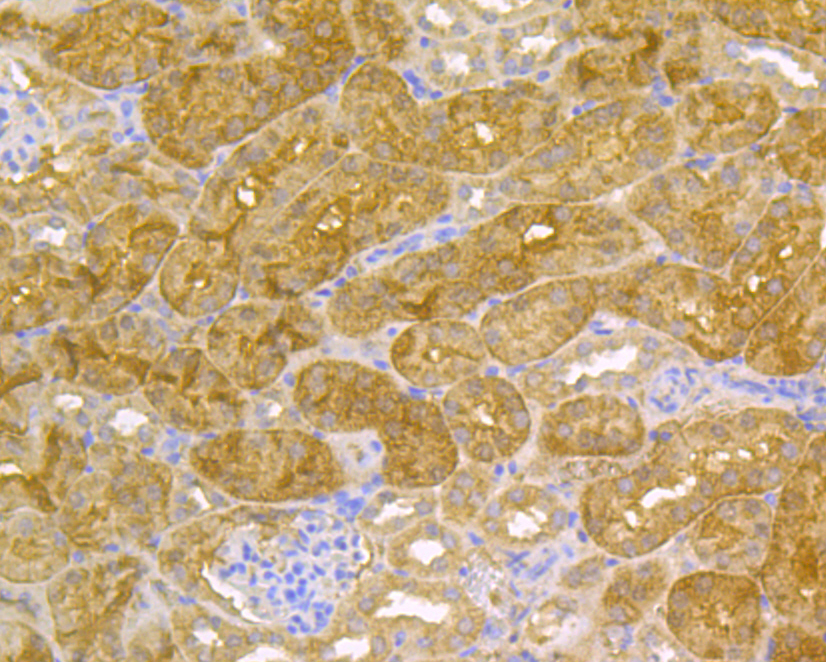
Fig5: Immunohistochemical analysis of paraffin-embedded mouse kidney tissue using anti-Methyltransferase-like 26 antibody. Counter stained with hematoxylin.Application
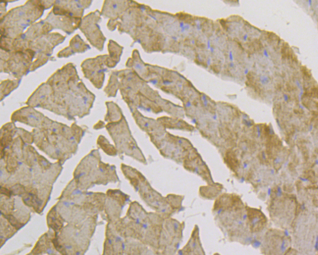
Fig6: Immunohistochemical analysis of paraffin-embedded mouse heart tissue using anti-Methyltransferase-like 26 antibody. Counter stained with hematoxylin.Application
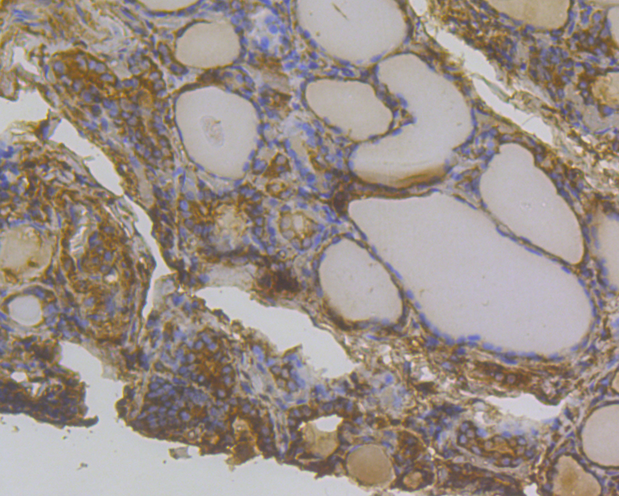
Fig7: Immunohistochemical analysis of paraffin-embedded human thyroid cancer tissue using anti-Methyltransferase-like 26 antibody. Counter stained with hematoxylin.Application
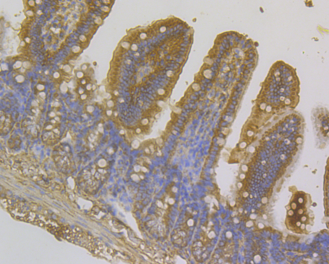
Fig8: Immunohistochemical analysis of paraffin-embedded mouse small intestine tissue using anti-Methyltransferase-like 26 antibody. Counter stained with hematoxylin.Application
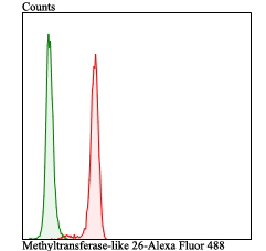
Fig9: Flow cytometric analysis of SH-SY-5Y cells with Methyltransferase-like 26 antibody at 1/100 dilution (red) compared with an unlabelled control (cells without incubation with primary antibody; green). Alexa Fluor 488-conjugated goat anti-rabbit IgG was used as the secondary antibody.| Product Name | Methyltransferase-like 26 |
|---|---|
| Antibody Type | Primary Antibodies |
| Product description | Also named Chromosome 16 open reading frame 13, also called C16orf13, is a protein-coding gene of unknown function, also known as JFP2. Though the function of this gene is unknown, various data have revealed that it is expressed at high levels in various cancerous tissues. Underexpression of this gene has also been linked to disease consequences in humans. |
| Immunogen | Synthetic peptide within human 45-70 aa. |
| Clonality | Polyclonal |
|---|---|
| Isotype | IgG |
| Host Species | Rabbit |
| Tested Applications | WBICCIHCFC |
| WB:1:500 ICC:1:50-1:200 IHC:1:50-1:200 FC:1:50-1:100 |
|
| Species Reactivity | HumanMouseRat |
| Concentration | 1mg/ml |
| Molecular Weight(MW) | 22 kDa |
|---|---|
| Cellular Localization | Cytoplasm. |
Application
Fig1: Western blot analysis of Methyltransferase-like 26 on mouse kidney tissue lysate using anti-Methyltransferase-like 26 antibody at 1/500 dilution.
Application
Fig2: ICC staining Methyltransferase-like 26 in SH-SY-5Y cells (green). The nuclear counter stain is DAPI (blue). Cells were fixed in paraformaldehyde, permeabilised with 0.25% Triton X100/PBS.
Application
Fig3: ICC staining Methyltransferase-like 26 in MCF-7 cells (green). The nuclear counter stain is DAPI (blue). Cells were fixed in paraformaldehyde, permeabilised with 0.25% Triton X100/PBS.
Application
Fig4: Immunohistochemical analysis of paraffin-embedded human lung tissue using anti-Methyltransferase-like 26 antibody. Counter stained with hematoxylin.
Application
Fig5: Immunohistochemical analysis of paraffin-embedded mouse kidney tissue using anti-Methyltransferase-like 26 antibody. Counter stained with hematoxylin.
Application
Fig6: Immunohistochemical analysis of paraffin-embedded mouse heart tissue using anti-Methyltransferase-like 26 antibody. Counter stained with hematoxylin.
Application
Fig7: Immunohistochemical analysis of paraffin-embedded human thyroid cancer tissue using anti-Methyltransferase-like 26 antibody. Counter stained with hematoxylin.
Application
Fig8: Immunohistochemical analysis of paraffin-embedded mouse small intestine tissue using anti-Methyltransferase-like 26 antibody. Counter stained with hematoxylin.
Application
Fig9: Flow cytometric analysis of SH-SY-5Y cells with Methyltransferase-like 26 antibody at 1/100 dilution (red) compared with an unlabelled control (cells without incubation with primary antibody; green). Alexa Fluor 488-conjugated goat anti-rabbit IgG was used as the secondary antibody.| Positive Control | Mouse kidney tissue, 293T, MCF-7, SH-SY-5Y, human lung tissue, human thyroid cancer tissue, mouse small intestine tissue, mouse heart tissue. |
|---|---|
| Application Notes | WB:1:500 ICC:1:50-1:200 IHC:1:50-1:200 FC:1:50-1:100 |
| Form | Liquid |
|---|---|
| Storage Instructions | Store at +4℃ after thawing. Aliquot store at -20℃. Avoid repeated freeze / thaw cycles. |
| Storage Buffer | 1*TBS (pH7.4), 0.5%BSA, 50%Glycerol. Preservative: 0.05% Sodium Azide. |


