Application
Fig1: Western blot analysis of Tmem248 on SkBr3 (1) and K562 cells lysates using anti-Tmem248 antibody.Application
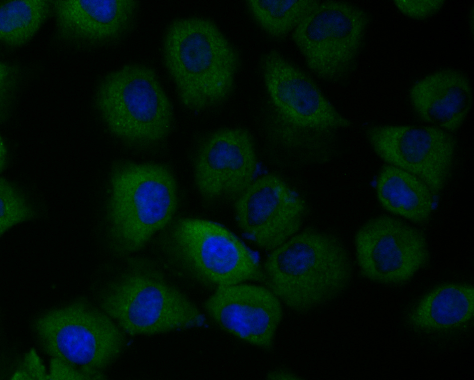
Fig2: ICC staining Tmem248 in HUVEC cells (green). The nuclear counter stain is DAPI (blue). Cells were fixed in paraformaldehyde, permeabilised with 0.25% Triton X100/PBS.Application
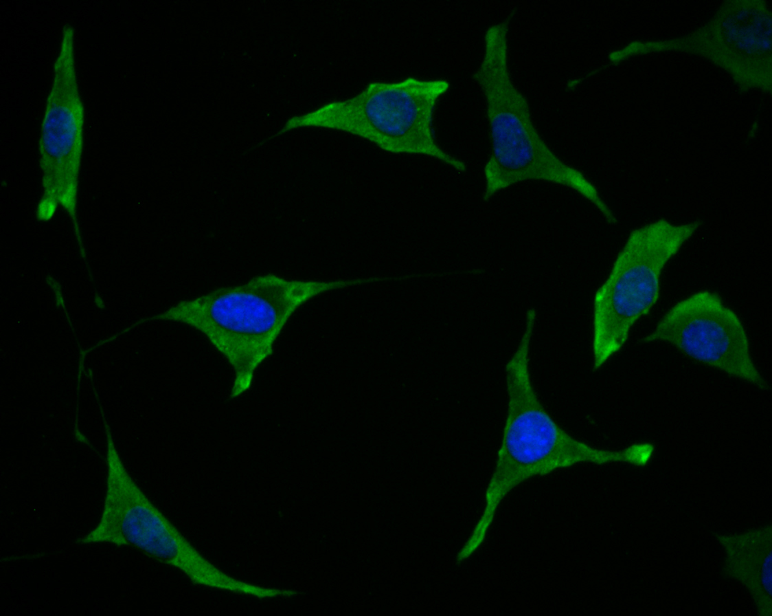
Fig3: ICC staining Tmem248 in SH-SY-5Y cells (green). The nuclear counter stain is DAPI (blue). Cells were fixed in paraformaldehyde, permeabilised with 0.25% Triton X100/PBS.Application
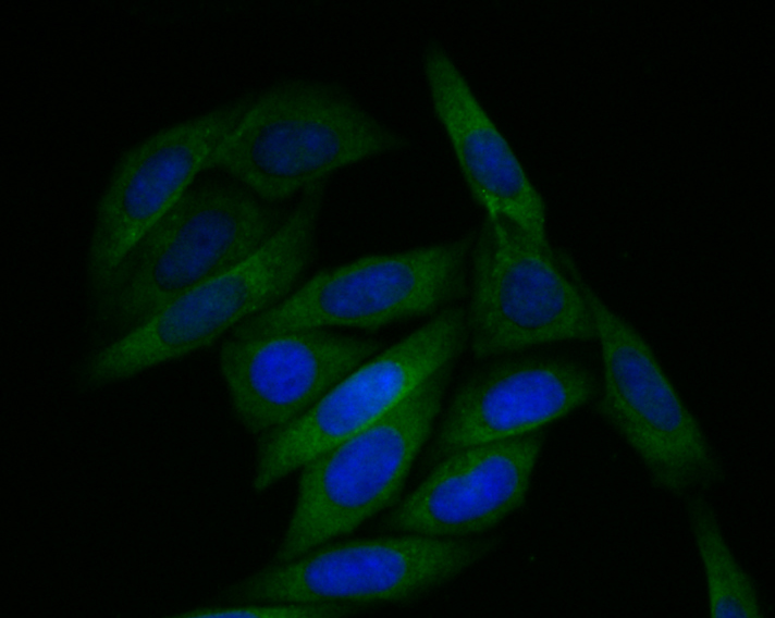
Fig4: ICC staining Tmem248 in SiHa cells (green). The nuclear counter stain is DAPI (blue). Cells were fixed in paraformaldehyde, permeabilised with 0.25% Triton X100/PBS.Application
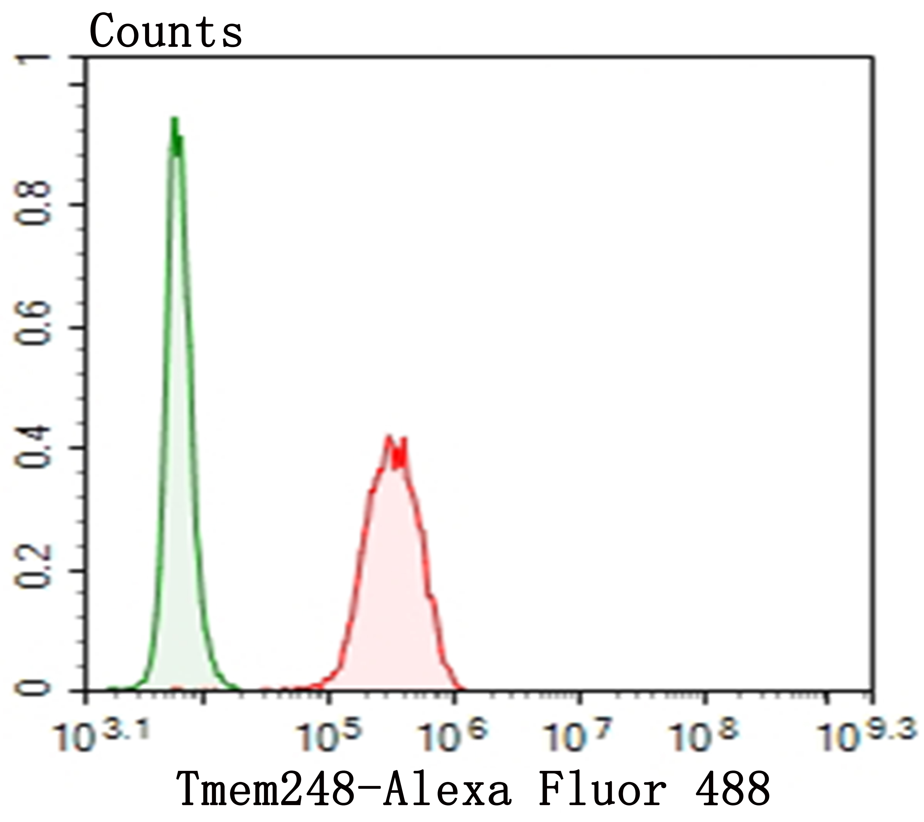
Fig5: Flow cytometric analysis of SH-SY-5Y cells with Tmem248 antibody at 1/100 dilution (red) compared with an unlabelled control (cells without incubation with primary antibody; green). Alexa Fluor 488-conjugated goat anti-rabbit IgG was used as the secondary antibody.| Product Name | Tmem248 |
|---|---|
| Antibody Type | Primary Antibodies |
| Product description | This gene is thought to be involved in apoptosis, and may also be involved in hematopoietic development and differentiation. The use of alternative splice sites and promotors result in multiple transcript variants encoding different isoforms. Promotes osteoblast cell differentiation and terminal mineralization. Plays a role in inducing the cell cycle arrest via inhibiting CCND1 expression in all-trans-retinoic acid (ATRA) signal pathway. |
| Immunogen | Synthetic peptide corresponding to C-terminal human Tmem248. |
| Clonality | Polyclonal |
|---|---|
| Isotype | IgG |
| Host Species | Rabbit |
| Tested Applications | WBICCFC |
| WB:1:500-1,000 ICC:1:50-1:200 FC:1:50-1:100 |
|
| Species Reactivity | HumanMouseRat |
| Concentration | 1mg/ml |
| Molecular Weight(MW) | 35/22 kDa (Predicted band size) |
|---|---|
| Cellular Localization | Membrane. |
Application
Fig1: Western blot analysis of Tmem248 on SkBr3 (1) and K562 cells lysates using anti-Tmem248 antibody.
Application
Fig2: ICC staining Tmem248 in HUVEC cells (green). The nuclear counter stain is DAPI (blue). Cells were fixed in paraformaldehyde, permeabilised with 0.25% Triton X100/PBS.
Application
Fig3: ICC staining Tmem248 in SH-SY-5Y cells (green). The nuclear counter stain is DAPI (blue). Cells were fixed in paraformaldehyde, permeabilised with 0.25% Triton X100/PBS.
Application
Fig4: ICC staining Tmem248 in SiHa cells (green). The nuclear counter stain is DAPI (blue). Cells were fixed in paraformaldehyde, permeabilised with 0.25% Triton X100/PBS.
Application
Fig5: Flow cytometric analysis of SH-SY-5Y cells with Tmem248 antibody at 1/100 dilution (red) compared with an unlabelled control (cells without incubation with primary antibody; green). Alexa Fluor 488-conjugated goat anti-rabbit IgG was used as the secondary antibody.| Positive Control | SkBr3, K562, SH-SY-5Y, HUVEC, SiHa. |
|---|---|
| Application Notes | WB:1:500-1,000 ICC:1:50-1:200 FC:1:50-1:100 |
| Form | Liquid |
|---|---|
| Storage Instructions | Store at +4℃ after thawing. Aliquot store at -20℃. Avoid repeated freeze / thaw cycles. |
| Storage Buffer | 1*TBS (pH7.4), 0.5%BSA, 50%Glycerol. Preservative: 0.05% Sodium Azide. |


