Application
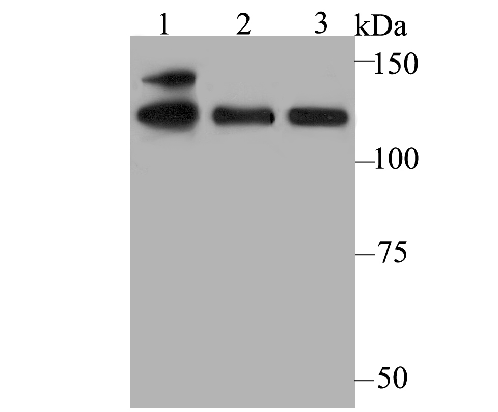
Fig1: Western blot analysis of USP28 on different cell lysates using anti-USP28 antibody at 1/500 dilution. Positive control: Lane1: K562 Lane2: A549 Lane3: DaudiApplication
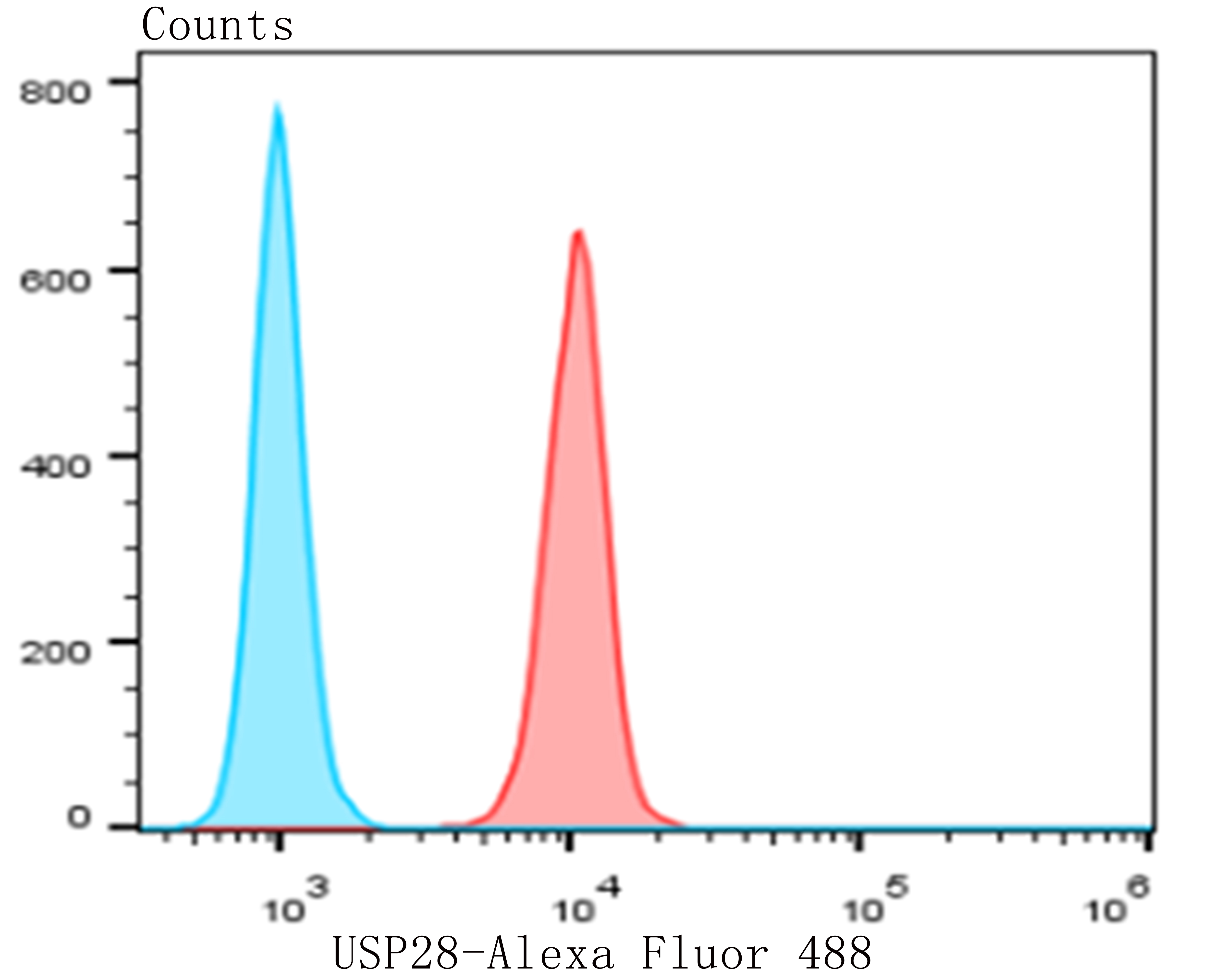
Fig2: Flow cytometric analysis of MG-63 cells with USP28 antibody at 1/50 dilution (red) compared with an unlabelled control (cells without incubation with primary antibody; blue). Alexa Fluor 488-conjugated goat anti-rabbit IgG was used as the secondary antibody.| Product Name | USP28 |
|---|---|
| Antibody Type | Primary Antibodies |
| Antigen Alias | Deubiquitinating enzyme 28 antibody KIAA1515 antibody Ubiquitin carboxyl terminal hydrolase 28 antibody Ubiquitin carboxyl-terminal hydrolase 28 antibody ubiquitin carboxyl-terminal hydrolase 28 variant 1 antibody Ubiquitin specific peptidase 28 antibody Ubiquitin specific processing protease 28 antibody Ubiquitin specific protease 28 antibody Ubiquitin thioesterase 28 antibody Ubiquitin thiolesterase 28 antibody Ubiquitin-specific-processing protease 28 antibody UBP28_HUMAN antibody USP 28 antibody USP28 antibody USP28 protein antibody |
| Product description | Deubiquitinase involved in DNA damage response checkpoint and MYC proto-oncogene stability. Involved in DNA damage induced apoptosis by specifically deubiquitinating proteins of the DNA damage pathway such as CLSPN. Also involved in G2 DNA damage checkpoint, by deubiquitinating CLSPN, and preventing its degradation by the anaphase promoting complex/cyclosome (APC/C). In contrast, it does not deubiquitinate PLK1. Specifically deubiquitinates MYC in the nucleoplasm, leading to prevent MYC degradation by the proteasome: acts by specifically interacting with isoform 1 of FBXW7 (FBW7alpha) in the nucleoplasm and counteracting ubiquitination of MYC by the SCF(FBW7) complex. In contrast, it does not interact with isoform 4 of FBXW7 (FBW7gamma) in the nucleolus, allowing MYC degradation and explaining the selective MYC degradation in the nucleolus. Deubiquitinates ZNF304, hence preventing ZNF304 degradation by the proteasome and leading to the activated KRAS-mediated promoter hypermethylation and transcriptional silencing of tumor suppressor genes (TSGs) in a subset of colorectal cancers (CRC) cells. |
| Immunogen | Recombinant protein within human USP28 aa 550-700. |
| Clonality | Polyclonal |
|---|---|
| Isotype | IgG |
| Host Species | Rabbit |
| Tested Applications | WBFC |
| WB:1:200-1:1,000 FC:1:50-1:100 |
|
| Species Reactivity | Human |
| Concentration | 1mg/ml |
| Alternative Names | Deubiquitinating enzyme 28 antibody KIAA1515 antibody Ubiquitin carboxyl terminal hydrolase 28 antibody Ubiquitin carboxyl-terminal hydrolase 28 antibody ubiquitin carboxyl-terminal hydrolase 28 variant 1 antibody Ubiquitin specific peptidase 28 antibody Ubiquitin specific processing protease 28 antibody Ubiquitin specific protease 28 antibody Ubiquitin thioesterase 28 antibody Ubiquitin thiolesterase 28 antibody Ubiquitin-specific-processing protease 28 antibody UBP28_HUMAN antibody USP 28 antibody USP28 antibody USP28 protein antibody |
|---|---|
| Molecular Weight(MW) | 122 kDa |
| Cellular Localization | Nucleus. |
| SwissProt ID | Q96RU2 |
|---|
Application
Fig1: Western blot analysis of USP28 on different cell lysates using anti-USP28 antibody at 1/500 dilution. Positive control: Lane1: K562 Lane2: A549 Lane3: Daudi
Application
Fig2: Flow cytometric analysis of MG-63 cells with USP28 antibody at 1/50 dilution (red) compared with an unlabelled control (cells without incubation with primary antibody; blue). Alexa Fluor 488-conjugated goat anti-rabbit IgG was used as the secondary antibody.| Positive Control | K562, A549, Daudi. |
|---|---|
| Application Notes | WB:1:200-1:1,000 FC:1:50-1:100 |
| Form | Liquid |
|---|---|
| Storage Instructions | Store at +4℃ after thawing. Aliquot store at -20℃. Avoid repeated freeze / thaw cycles. |
| Storage Buffer | 1*TBS (pH7.4), 0.5%BSA, 50%Glycerol. Preservative: 0.05% Sodium Azide. |


