Application
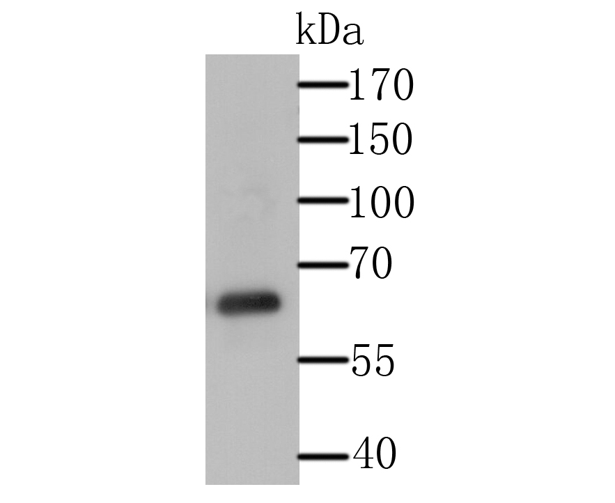
Fig1: Western blot analysis of USP14 on Daudi cell lysate using anti-USP14 antibody at 1/500 dilution.Application
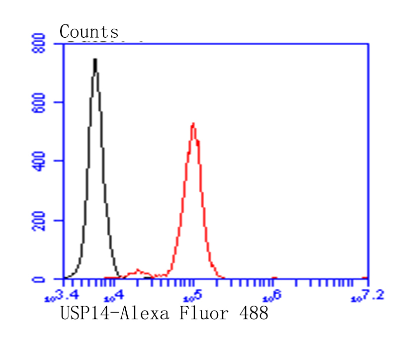
Fig2: Flow cytometric analysis of SiHa cells with USP14 antibody at 1/100 dilution (red) compared with an unlabelled control (cells without incubation with primary antibody;black). Alexa Fluor 488-conjugated goat anti-rabbit IgG was used as the secondary antibody.| Product Name | USP14 |
|---|---|
| Antibody Type | Primary Antibodies |
| Antigen Alias | Deubiquitinating enzyme 14 antibody TGT antibody tRNA guanine transglycosylase 60 kD subunit antibody tRNA guanine transglycosylase antibody Ubiquitin carboxyl terminal hydrolase 14 antibody Ubiquitin carboxyl-terminal hydrolase 14 antibody Ubiquitin specific peptidase 14 antibody Ubiquitin specific processing protease 14 antibody Ubiquitin specific protease 14 antibody Ubiquitin thiolesterase 14 antibody Ubiquitin-specific-processing protease 14 antibody UBP14_HUMAN antibody USP 14 antibody USP14 antibody |
| Product description | Proteasome-associated deubiquitinase which releases ubiquitin from the proteasome targeted ubiquitinated proteins. Ensures the regeneration of ubiquitin at the proteasome. Is a reversibly associated subunit of the proteasome and a large fraction of proteasome-free protein exists within the cell. Required for the degradation of the chemokine receptor CXCR4 which is critical for CXCL12-induced cell chemotaxis. Serves also as a physiological inhibitor of endoplasmic reticulum-associated degradation under the non-stressed condition by inhibiting the degradation of unfolded endoplasmic reticulum proteins via interaction with ERN1. Indispensable for synaptic development and function at neuromuscular junctions. Plays a role in the innate immune defense against viruses by stabilizing the viral DNA sensor CGAS and thus inhibiting its autophagic degradation. |
| Immunogen | Recombinant protein within human USP14 aa 200-450. |
| Clonality | Polyclonal |
|---|---|
| Isotype | IgG |
| Host Species | Rabbit |
| Tested Applications | WBFC |
| WB:1:500-1:1,000 FC: 1:50-1:100 |
|
| Species Reactivity | Human |
| Concentration | 1mg/ml |
| Alternative Names | Deubiquitinating enzyme 14 antibody TGT antibody tRNA guanine transglycosylase 60 kD subunit antibody tRNA guanine transglycosylase antibody Ubiquitin carboxyl terminal hydrolase 14 antibody Ubiquitin carboxyl-terminal hydrolase 14 antibody Ubiquitin specific peptidase 14 antibody Ubiquitin specific processing protease 14 antibody Ubiquitin specific protease 14 antibody Ubiquitin thiolesterase 14 antibody Ubiquitin-specific-processing protease 14 antibody UBP14_HUMAN antibody USP 14 antibody USP14 antibody |
|---|---|
| Molecular Weight(MW) | 56 kDa |
| Cellular Localization | Plasma membrane. |
| SwissProt ID | P54578 |
|---|
Application
Fig1: Western blot analysis of USP14 on Daudi cell lysate using anti-USP14 antibody at 1/500 dilution.
Application
Fig2: Flow cytometric analysis of SiHa cells with USP14 antibody at 1/100 dilution (red) compared with an unlabelled control (cells without incubation with primary antibody;black). Alexa Fluor 488-conjugated goat anti-rabbit IgG was used as the secondary antibody.| Positive Control | Daudi, SiHa. |
|---|---|
| Application Notes | WB:1:500-1:1,000 FC: 1:50-1:100 |
| Form | Liquid |
|---|---|
| Storage Instructions | Store at +4℃ after thawing. Aliquot store at -20℃. Avoid repeated freeze / thaw cycles. |
| Storage Buffer | 1*TBS (pH7.4), 0.5%BSA, 50%Glycerol. Preservative: 0.05% Sodium Azide. |


