Application
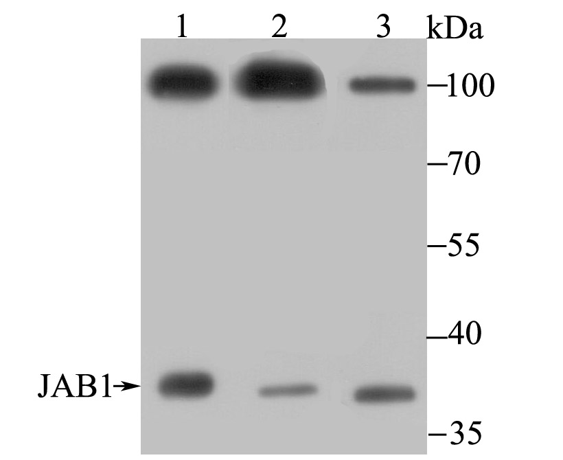
Fig1: Western blot analysis of JAB1 on different lysate using anti-JAB1 antibody at 1/500 dilution. Positive control: Lane 1: Mouse testis tissue Lane 2: PC-3M Lane 3: A549Application
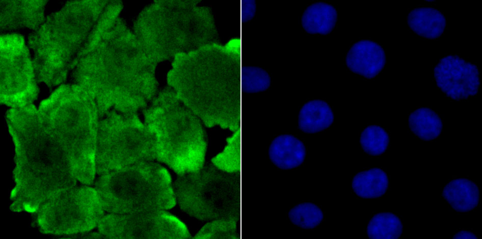
Fig2: ICC staining JAB1 in A549 cells (green). The nuclear counter stain is DAPI (blue). Cells were fixed in paraformaldehyde, permeabilised with 0.25% Triton X100/PBS.Application
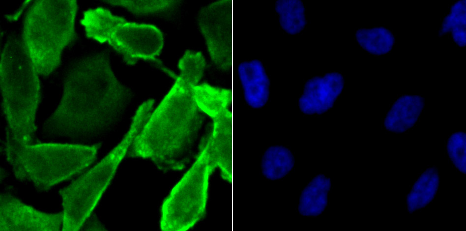
Fig3: ICC staining JAB1 in PC-3M cells (green). The nuclear counter stain is DAPI (blue). Cells were fixed in paraformaldehyde, permeabilised with 0.25% Triton X100/PBS.Application
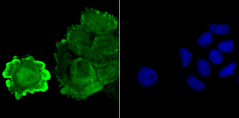
Fig4: ICC staining JAB1 in A431 cells (green). The nuclear counter stain is DAPI (blue). Cells were fixed in paraformaldehyde, permeabilised with 0.25% Triton X100/PBS.Application
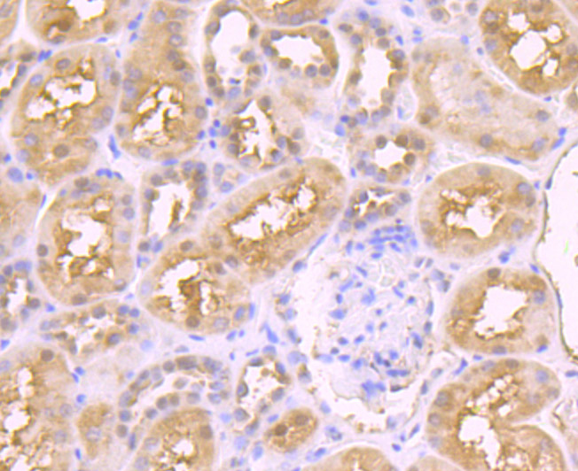
Fig5: Immunohistochemical analysis of paraffin-embedded human kidney tissue using anti-JAB1 antibody. Counter stained with hematoxylin.Application
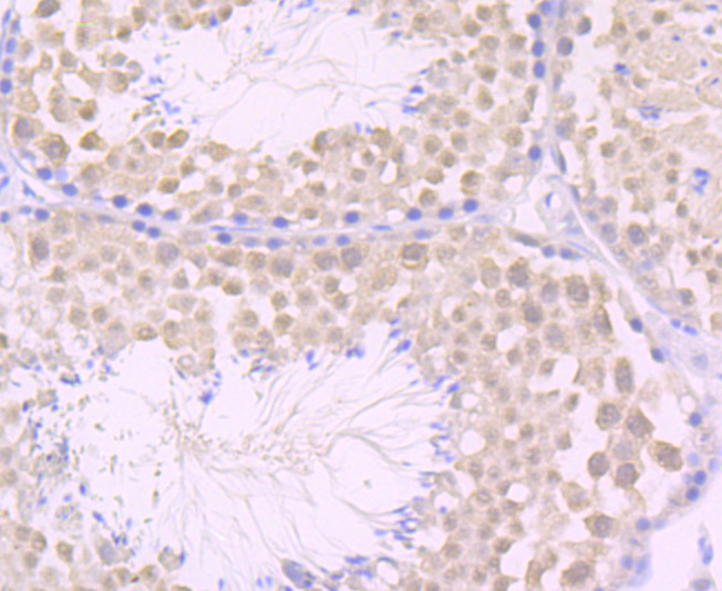
Fig6: Immunohistochemical analysis of paraffin-embedded mouse testis tissue using anti-JAB1 antibody. Counter stained with hematoxylin.Application
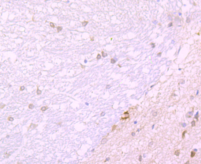
Fig7: Immunohistochemical analysis of paraffin-embedded rat brain tissue using anti-JAB1 antibody. Counter stained with hematoxylin.| Product Name | JAB1 [JB34-96] |
|---|---|
| Antibody Type | Primary Antibodies |
| Antigen Alias | 38 kDa Mov34 homolog antibody COP9 (constitutive photomorphogenic) homolog subunit 5 antibody COP9 constitutive photomorphogenic homolog subunit 5 antibody COP9 signalosome complex subunit 5 antibody COP9 signalosome subunit 5 antibody Cop9 subunit 5 antibody COPS 5 antibody cops5 antibody CSN 5 antibody CSN5 antibody CSN5_HUMAN antibody JAB 1 antibody Jun activation domain binding protein 1 antibody Jun activation domain binding protein antibody Jun activation domain-binding protein 1 antibody MGC3149 antibody MOV 34 antibody MOV34 antibody MOV34 family, 38-KD member antibody SGN 5 antibody SGN5 antibody Signalosome subunit 5 antibody |
| Product description | Genes belonging to the Jun and Fos oncogene families encode nuclear proteins that are found to be associated with a number of transcriptional complexes. The c-Jun protein is a major component of the transcription factor AP-1, originally shown to mediate phorbol ester tumor promoter (TPA)-induced expression of responsive genes through the TPA-response element (TRE). The Jun proteins form homo- and heterodimers which bind the TRE, but the Fos proteins are active only as heterodimers with any of the Jun proteins. Fos/Jun heterodimers have a much higher affinity for the TRE than Jun homodimers. Ha-Ras augments c-Jun activity and stimulates phosphorylation of its activation domain. The coactivator of Jun, designated JAB1 (for Jun-activation domain-binding protein), interacts with c-Jun and Jun D, but not with Jun B or v-Jun. This interaction enhances the transactivating ability of Jun proteins by stabilizing their binding to the TRE. The gene encoding JAB1 maps to human chromosome 8q12.3. |
| Immunogen | Recombinant protein |
| Clonality | Monoclonal |
|---|---|
| Isotype | IgG |
| Host Species | Recombinant rabbit |
| Tested Applications | WBICCIFIHCIP |
| WB:1:500 ICC:1:100-1:500 IHC:1:50-1:200 |
|
| Species Reactivity | HumanMouseRat |
| Concentration | 1mg/ml |
| Alternative Names | 38 kDa Mov34 homolog antibody
COP9 (constitutive photomorphogenic) homolog subunit 5 antibody
COP9 constitutive photomorphogenic homolog subunit 5 antibody
COP9 signalosome complex subunit 5 antibody
COP9 signalosome subunit 5 antibody
Cop9 subunit 5 antibody
COPS 5 antibody
cops5 antibody
CSN 5 antibody
CSN5 antibody
CSN5_HUMAN antibody
JAB 1 antibody
Jun activation domain binding protein 1 antibody
Jun activation domain binding protein antibody
Jun activation domain-binding protein 1 antibody
MGC3149 antibody
MOV 34 antibody
MOV34 antibody
MOV34 family 38-KD member antibody SGN 5 antibody SGN5 antibody Signalosome subunit 5 antibody |
|---|---|
| Molecular Weight(MW) | 37 kDa, additional band 100 kDa. |
| Cellular Localization | Cytoplasm. Nucleus. |
Application
Fig1: Western blot analysis of JAB1 on different lysate using anti-JAB1 antibody at 1/500 dilution. Positive control: Lane 1: Mouse testis tissue Lane 2: PC-3M Lane 3: A549
Application
Fig2: ICC staining JAB1 in A549 cells (green). The nuclear counter stain is DAPI (blue). Cells were fixed in paraformaldehyde, permeabilised with 0.25% Triton X100/PBS.
Application
Fig3: ICC staining JAB1 in PC-3M cells (green). The nuclear counter stain is DAPI (blue). Cells were fixed in paraformaldehyde, permeabilised with 0.25% Triton X100/PBS.
Application
Fig4: ICC staining JAB1 in A431 cells (green). The nuclear counter stain is DAPI (blue). Cells were fixed in paraformaldehyde, permeabilised with 0.25% Triton X100/PBS.
Application
Fig5: Immunohistochemical analysis of paraffin-embedded human kidney tissue using anti-JAB1 antibody. Counter stained with hematoxylin.
Application
Fig6: Immunohistochemical analysis of paraffin-embedded mouse testis tissue using anti-JAB1 antibody. Counter stained with hematoxylin.
Application
Fig7: Immunohistochemical analysis of paraffin-embedded rat brain tissue using anti-JAB1 antibody. Counter stained with hematoxylin.| Positive Control | A431, A549, PC-3M, rat brain tissue, human kidney tissue, mouse testis tissue. |
|---|---|
| Application Notes | WB:1:500 ICC:1:100-1:500 IHC:1:50-1:200 |
| Form | Liquid |
|---|---|
| Storage Instructions | Store at +4℃ after thawing. Aliquot store at -20℃ or -80℃. Avoid repeated freeze / thaw cycles. |
| Storage Buffer | 1*TBS (pH7.4), 1%BSA, 40%Glycerol. Preservative: 0.05% Sodium Azide. |


