Application
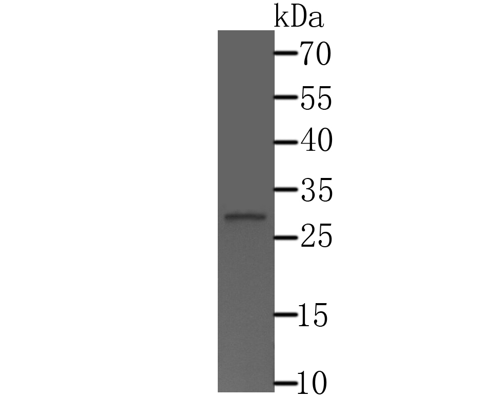
Fig1: Western blot analysis of CTDSPL on HepG2 cell lysate using anti-CTDSPL antibody at 1/500 dilution.Application
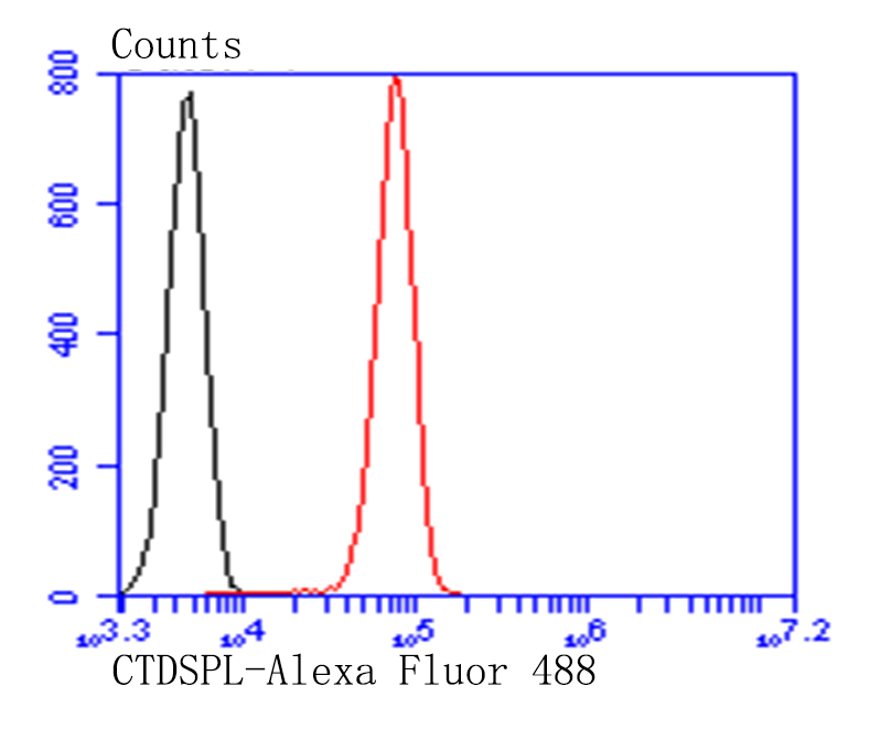
Fig2: Flow cytometric analysis of HepG2 cells with CFTR antibody at 1/100 dilution (red) compared with an unlabelled control (cells without incubation with primary antibody; black). Alexa Fluor 488-conjugated goat anti-rabbit IgG was used as the secondary antibody.| Product Name | CTDSPL |
|---|---|
| Antibody Type | Primary Antibodies |
| Antigen Alias | C3orf8 antibody Carboxy-terminal domain RNA polymerase II polypeptide A small phosphatase 3 antibody CTD (carboxy-terminal domain, RNA polymerase II, polypeptide A) small phosphatase-like antibody CTD small phosphatase-like protein antibody CTDSL_HUMAN antibody CTDSP-like antibody Ctdspl antibody hYA22 antibody N-like protein antibody NIF-like protein antibody NLI-interacting factor 1 antibody Nuclear LIM interactor-interacting factor 1 antibody Protein YA22 antibody RB protein serine phosphatase from chromosome 3 antibody RBSP3 antibody SCP3 antibody Small C-terminal domain phosphatase 3 antibody Small CTD phosphatase 3 antibody |
| Product description | Recruited by REST to neuronal genes that contain RE-1 elements, leading to neuronal gene silencing in non-neuronal cells (By similarity). Preferentially catalyzes the dephosphorylation of 'Ser-5' within the tandem 7 residue repeats in the C-terminal domain (CTD) of the largest RNA polymerase II subunit POLR2A. Negatively regulates RNA polymerase II transcription, possibly by controlling the transition from initiation/capping to processive transcript elongation. |
| Immunogen | Recombinant protein within human CTDSPL aa 50-300. |
| Clonality | Polyclonal |
|---|---|
| Isotype | IgG |
| Host Species | Rabbit |
| Tested Applications | WBFC |
| WB:1:500 FC: 1:50-1:100 |
|
| Species Reactivity | HumanMouseRat |
| Concentration | 1mg/ml |
| Alternative Names | C3orf8 antibody
Carboxy-terminal domain RNA polymerase II polypeptide A small phosphatase 3 antibody
CTD (carboxy-terminal domain RNA polymerase II polypeptide A) small phosphatase-like antibody CTD small phosphatase-like protein antibody CTDSL_HUMAN antibody CTDSP-like antibody Ctdspl antibody hYA22 antibody N-like protein antibody NIF-like protein antibody NLI-interacting factor 1 antibody Nuclear LIM interactor-interacting factor 1 antibody Protein YA22 antibody RB protein serine phosphatase from chromosome 3 antibody RBSP3 antibody SCP3 antibody Small C-terminal domain phosphatase 3 antibody Small CTD phosphatase 3 antibody |
|---|---|
| Molecular Weight(MW) | 31kDa |
| Cellular Localization | Nucleus. |
Application
Fig1: Western blot analysis of CTDSPL on HepG2 cell lysate using anti-CTDSPL antibody at 1/500 dilution.
Application
Fig2: Flow cytometric analysis of HepG2 cells with CFTR antibody at 1/100 dilution (red) compared with an unlabelled control (cells without incubation with primary antibody; black). Alexa Fluor 488-conjugated goat anti-rabbit IgG was used as the secondary antibody.| Positive Control | HepG2. |
|---|---|
| Application Notes | WB:1:500 FC: 1:50-1:100 |
| Form | Liquid |
|---|---|
| Storage Instructions | Store at +4℃ after thawing. Aliquot store at -20℃. Avoid repeated freeze / thaw cycles. |
| Storage Buffer | 1*TBS (pH7.4), 0.5%BSA, 50%Glycerol. Preservative: 0.05% Sodium Azide. |


