Application
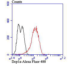
Fig7: Flow cytometric analysis of LOVO cells with Dcp1a antibody at 1/100 dilution (red) compared with an unlabelled control (cells without incubation with primary antibody; black). Alexa Fluor 488-conjugated goat anti-rabbit IgG was used as the secondary antibody.Application
Fig7: Flow cytometric analysis of LOVO cells with Dcp1a antibody at 1/100 dilution (red) compared with an unlabelled control (cells without incubation with primary antibody; black). Alexa Fluor 488-conjugated goat anti-rabbit IgG was used as the secondary antibody.Application
Fig7: Flow cytometric analysis of LOVO cells with Dcp1a antibody at 1/100 dilution (red) compared with an unlabelled control (cells without incubation with primary antibody; black). Alexa Fluor 488-conjugated goat anti-rabbit IgG was used as the secondary antibody.Application
Fig7: Flow cytometric analysis of LOVO cells with Dcp1a antibody at 1/100 dilution (red) compared with an unlabelled control (cells without incubation with primary antibody; black). Alexa Fluor 488-conjugated goat anti-rabbit IgG was used as the secondary antibody.Application
Fig7: Flow cytometric analysis of LOVO cells with Dcp1a antibody at 1/100 dilution (red) compared with an unlabelled control (cells without incubation with primary antibody; black). Alexa Fluor 488-conjugated goat anti-rabbit IgG was used as the secondary antibody.Application
Fig7: Flow cytometric analysis of LOVO cells with Dcp1a antibody at 1/100 dilution (red) compared with an unlabelled control (cells without incubation with primary antibody; black). Alexa Fluor 488-conjugated goat anti-rabbit IgG was used as the secondary antibody.Application
Fig7: Flow cytometric analysis of LOVO cells with Dcp1a antibody at 1/100 dilution (red) compared with an unlabelled control (cells without incubation with primary antibody; black). Alexa Fluor 488-conjugated goat anti-rabbit IgG was used as the secondary antibody.| Product Name | Dcp1a [JB51-34] |
|---|---|
| Antibody Type | Primary Antibodies |
| Antigen Alias | DCP1 decapping enzyme homolog A antibody Dcp1a antibody DCP1A_HUMAN antibody Decapping enzyme hDcp1a antibody Decapping mRNA 1A antibody HSA275986 antibody mRNA decapping enzyme 1A antibody mRNA-decapping enzyme 1A antibody Nbla00360 antibody Putative protein product of Nbla00360 antibody Smad4 interacting transcriptional co activator antibody Smad4-interacting transcriptional co-activator antibody Smad4-interacting transcriptional co-activator antibody SMAD4IP1 antibody SMIF antibody Transcription factor SMIF antibody |
| Product description | Cleavage of the 5'-cap structure is involved in the major 5'-to-3' and nonsense-mediated mRNA decay pathways. The protein complex consisting of Dcp1 and Dcp2 has been identified as the species responsible for the decapping reaction in Saccharomyces cerevisiae. In nonsense-mediated decay, the human decapping complex, made up of S. cerevisiae homologs Dcp1a and hDcp2, may be recruited to mRNAs containing premature termination codons by nonsense-mediated decay factor (Upf) proteins. hDcp2 specifically hydrolyzes methylated capped RNA to release m(7)GDP, thereby aiding in mRNA degradation. Both Dcp1a and hDcp2 colocalize in the cytoplasm. In addition, Dcp1a interacts with Smad4 forming a complex with TGFβ and BMP-4. Dcp1a and Smad4 interact directly through a EVH1/WH1 domain on Dcp1a and a proline-rich activation domain on Smad4. Smad4 is essential to nuclear translocation of Dcp1a as deletion of the Smad4-interacting domain (located in the N-terminal 100 amino acids) of Dcp1a eliminates TGFβ-induced nuclear translocation of Dcp1a. |
| Immunogen | Recombinant protein within C terminal Human Dcp1a. |
| Clonality | Monoclonal |
|---|---|
| Isotype | IgG |
| Host Species | Recombinant rabbit |
| Tested Applications | WBICCIHCFC |
| WB:1:500-1:2000 ICC:1:50-1:200 IHC:1:50-1:200 FC:1:50-1:100 |
|
| Species Reactivity | Human |
| Concentration | 1mg/ml |
| Alternative Names | DCP1 decapping enzyme homolog A antibody Dcp1a antibody DCP1A_HUMAN antibody Decapping enzyme hDcp1a antibody Decapping mRNA 1A antibody HSA275986 antibody mRNA decapping enzyme 1A antibody mRNA-decapping enzyme 1A antibody Nbla00360 antibody Putative protein product of Nbla00360 antibody Smad4 interacting transcriptional co activator antibody Smad4-interacting transcriptional co-activator antibody Smad4-interacting transcriptional co-activator antibody SMAD4IP1 antibody SMIF antibody Transcription factor SMIF antibody |
|---|---|
| Molecular Weight(MW) | 63 kDa(Predicted band size) |
| Cellular Localization | Cytoplasm. Nucleus. |
| SwissProt ID | Q9NPI6 |
|---|
Application
Fig7: Flow cytometric analysis of LOVO cells with Dcp1a antibody at 1/100 dilution (red) compared with an unlabelled control (cells without incubation with primary antibody; black). Alexa Fluor 488-conjugated goat anti-rabbit IgG was used as the secondary antibody.
Application
Fig7: Flow cytometric analysis of LOVO cells with Dcp1a antibody at 1/100 dilution (red) compared with an unlabelled control (cells without incubation with primary antibody; black). Alexa Fluor 488-conjugated goat anti-rabbit IgG was used as the secondary antibody.
Application
Fig7: Flow cytometric analysis of LOVO cells with Dcp1a antibody at 1/100 dilution (red) compared with an unlabelled control (cells without incubation with primary antibody; black). Alexa Fluor 488-conjugated goat anti-rabbit IgG was used as the secondary antibody.
Application
Fig7: Flow cytometric analysis of LOVO cells with Dcp1a antibody at 1/100 dilution (red) compared with an unlabelled control (cells without incubation with primary antibody; black). Alexa Fluor 488-conjugated goat anti-rabbit IgG was used as the secondary antibody.
Application
Fig7: Flow cytometric analysis of LOVO cells with Dcp1a antibody at 1/100 dilution (red) compared with an unlabelled control (cells without incubation with primary antibody; black). Alexa Fluor 488-conjugated goat anti-rabbit IgG was used as the secondary antibody.
Application
Fig7: Flow cytometric analysis of LOVO cells with Dcp1a antibody at 1/100 dilution (red) compared with an unlabelled control (cells without incubation with primary antibody; black). Alexa Fluor 488-conjugated goat anti-rabbit IgG was used as the secondary antibody.
Application
Fig7: Flow cytometric analysis of LOVO cells with Dcp1a antibody at 1/100 dilution (red) compared with an unlabelled control (cells without incubation with primary antibody; black). Alexa Fluor 488-conjugated goat anti-rabbit IgG was used as the secondary antibody.| Positive Control | LOVO, SH-SY-5Y, SiHa, human liver tissue, human placenta tissue. |
|---|---|
| Application Notes | WB:1:500-1:2000 ICC:1:50-1:200 IHC:1:50-1:200 FC:1:50-1:100 |
| Form | Liquid |
|---|---|
| Storage Instructions | Store at +4℃ after thawing. Aliquot store at -20℃. Avoid repeated freeze / thaw cycles. |
| Storage Buffer | 1*TBS (pH7.4), 1%BSA, 40%Glycerol. Preservative: 0.05% Sodium Azide. |


