Application
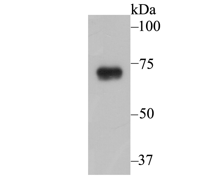
Fig1: Western blot analysis of SCNN1G on A431 cell lysate using anti-SCNN1G antibody at 1/1,000 dilution.Application
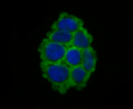
Fig2: ICC staining SCNN1G in 293T cells (green). The nuclear counter stain is DAPI (blue). Cells were fixed in paraformaldehyde, permeabilised with 0.25% Triton X100/PBS.Application
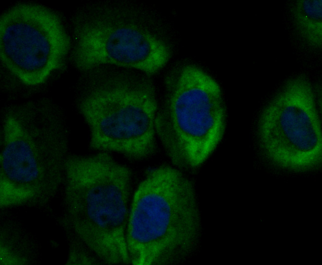
Fig3: ICC staining SCNN1G in A431 cells (green). The nuclear counter stain is DAPI (blue). Cells were fixed in paraformaldehyde, permeabilised with 0.25% Triton X100/PBS.Application
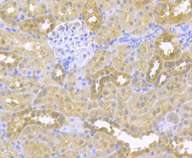
Fig4: Immunohistochemical analysis of paraffin-embedded rat kidney tissue using anti-SCNN1G antibody. Counter stained with hematoxylin.Application
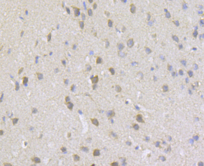
Fig5: Immunohistochemical analysis of paraffin-embedded mouse brain tissue using anti-SCNN1G antibody. Counter stained with hematoxylin.Application
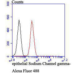
Fig6: Flow cytometric analysis of 293T cells with SCNN1G antibody at 1/100 dilution (red) compared with an unlabelled control (cells without incubation with primary antibody; black). Alexa Fluor 488-conjugated goat anti-rabbit IgG was used as the secondary antibody.| Product Name | SCNN1G |
|---|---|
| Antibody Type | Primary Antibodies |
| Antigen Alias | Amiloride sensitive epithelial sodium channel gamma subunit antibody Amiloride sensitive sodium channel subunit gamma antibody Amiloride-sensitive sodium channel subunit gamma antibody BESC3 antibody ENaC gamma subunit antibody ENaCG antibody ENaCgamma antibody Epithelial Na(+) channel subunit gamma antibody Epithelial Na+ channel subunit gamma antibody Gamma ENaC antibody Gamma NaCH antibody Gamma-ENaC antibody Gamma-NaCH antibody Nonvoltage gated sodium channel 1 subunit gamma antibody Nonvoltage-gated sodium channel 1 subunit gamma antibody PHA 1 antibody PHA1 antibody SCNEG antibody SCNN 1G antibody SCNN1G antibody SCNNG_HUMAN antibody Sodium channel epithelial 1 gamma subunit antibody Sodium channel non voltage gated 1 gamma subunit antibody Sodium channel nonvoltage gated 1 gamma antibody |
| Product description | Sodium permeable non-voltage-sensitive ion channel inhibited by the diuretic amiloride. Mediates the electrodiffusion of the luminal sodium (and water, which follows osmotically) through the apical membrane of epithelial cells. Plays an essential role in electrolyte and blood pressure homeostasis, but also in airway surface liquid homeostasis, which is important for proper clearance of mucus.; Controls the reabsorption of sodium in kidney, colon, lung and sweat glands. Also plays a role in taste perception. |
| Immunogen | Recombinant protein within human SCNN1G aa 250-350. |
| Clonality | Polyclonal |
|---|---|
| Isotype | IgG |
| Host Species | Rabbit |
| Tested Applications | WBICCIHCFC |
| WB:1:500-1:2,000 ICC:1:50-1:200 IHC:1:50-1:200 FC:1:50-1:100 |
|
| Species Reactivity | HumanMouseRat |
| Concentration | 1mg/ml |
| Alternative Names | Amiloride sensitive epithelial sodium channel gamma subunit antibody Amiloride sensitive sodium channel subunit gamma antibody Amiloride-sensitive sodium channel subunit gamma antibody BESC3 antibody ENaC gamma subunit antibody ENaCG antibody ENaCgamma antibody Epithelial Na(+) channel subunit gamma antibody Epithelial Na+ channel subunit gamma antibody Gamma ENaC antibody Gamma NaCH antibody Gamma-ENaC antibody Gamma-NaCH antibody Nonvoltage gated sodium channel 1 subunit gamma antibody Nonvoltage-gated sodium channel 1 subunit gamma antibody PHA 1 antibody PHA1 antibody SCNEG antibody SCNN 1G antibody SCNN1G antibody SCNNG_HUMAN antibody Sodium channel epithelial 1 gamma subunit antibody Sodium channel non voltage gated 1 gamma subunit antibody Sodium channel nonvoltage gated 1 gamma antibody |
|---|---|
| Molecular Weight(MW) | 74 kDa |
| Cellular Localization | Plasma membrane. |
Application
Fig1: Western blot analysis of SCNN1G on A431 cell lysate using anti-SCNN1G antibody at 1/1,000 dilution.
Application
Fig2: ICC staining SCNN1G in 293T cells (green). The nuclear counter stain is DAPI (blue). Cells were fixed in paraformaldehyde, permeabilised with 0.25% Triton X100/PBS.
Application
Fig3: ICC staining SCNN1G in A431 cells (green). The nuclear counter stain is DAPI (blue). Cells were fixed in paraformaldehyde, permeabilised with 0.25% Triton X100/PBS.
Application
Fig4: Immunohistochemical analysis of paraffin-embedded rat kidney tissue using anti-SCNN1G antibody. Counter stained with hematoxylin.
Application
Fig5: Immunohistochemical analysis of paraffin-embedded mouse brain tissue using anti-SCNN1G antibody. Counter stained with hematoxylin.
Application
Fig6: Flow cytometric analysis of 293T cells with SCNN1G antibody at 1/100 dilution (red) compared with an unlabelled control (cells without incubation with primary antibody; black). Alexa Fluor 488-conjugated goat anti-rabbit IgG was used as the secondary antibody.| Positive Control | A431, 293T, rat kidney tissue, mouse brain tissue. |
|---|---|
| Application Notes | WB:1:500-1:2,000 ICC:1:50-1:200 IHC:1:50-1:200 FC:1:50-1:100 |
| Form | Liquid |
|---|---|
| Storage Instructions | Store at +4℃ after thawing. Aliquot store at -20℃. Avoid repeated freeze / thaw cycles. |
| Storage Buffer | 1*TBS (pH7.4), 0.5%BSA, 50%Glycerol. Preservative: 0.05% Sodium Azide. |


