Application
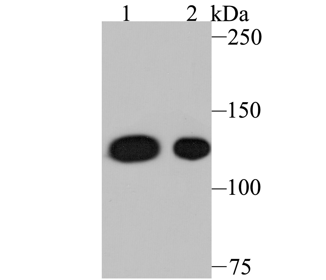
Fig1: Western blot analysis of Nesprin 1 on different lysates using anti-Nesprin 1 antibody at 1/500 dilution. Positive control: Lane 1: A549 Lane 2: Mouse spleen tissueApplication
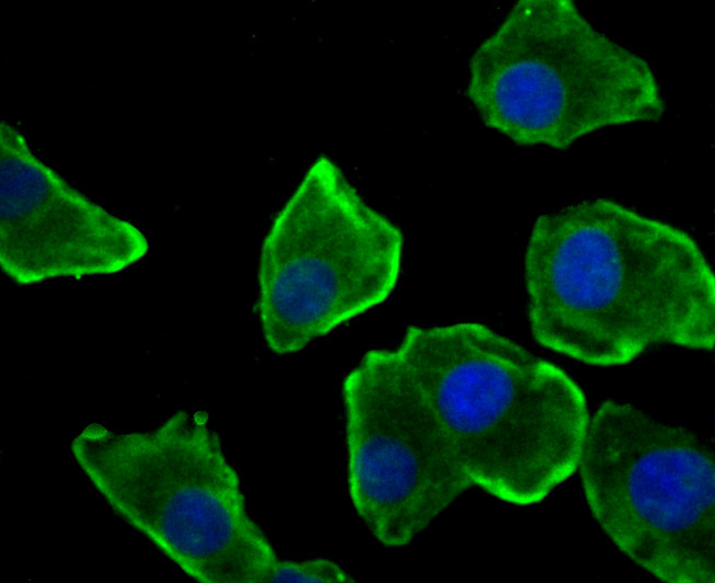
Fig2: ICC staining Nesprin 1 in A549 cells (green). The nuclear counter stain is DAPI (blue). Cells were fixed in paraformaldehyde, permeabilised with 0.25% Triton X100/PBS.Application
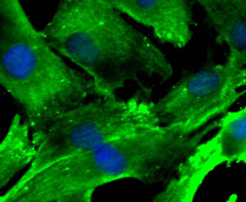
Fig3: ICC staining Nesprin 1 in C2C12 cells (green). The nuclear counter stain is DAPI (blue). Cells were fixed in paraformaldehyde, permeabilised with 0.25% Triton X100/PBS.Application
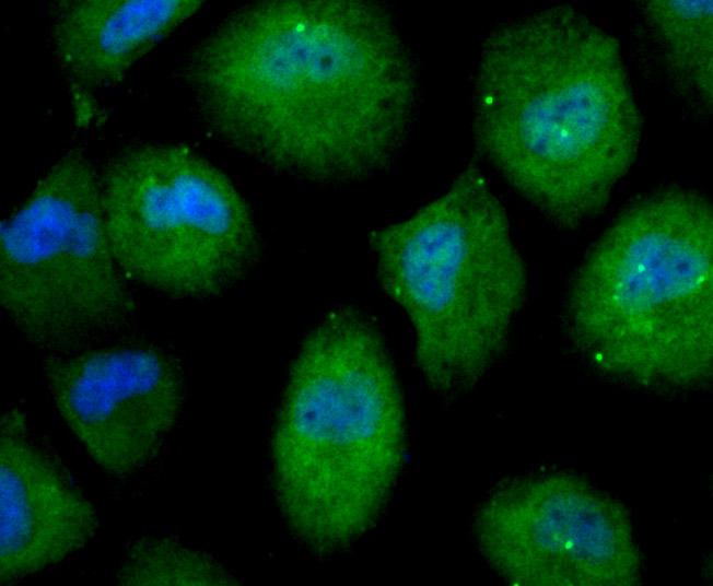
Fig4: ICC staining Nesprin 1 in HUVEC cells (green). The nuclear counter stain is DAPI (blue). Cells were fixed in paraformaldehyde, permeabilised with 0.25% Triton X100/PBS.Application
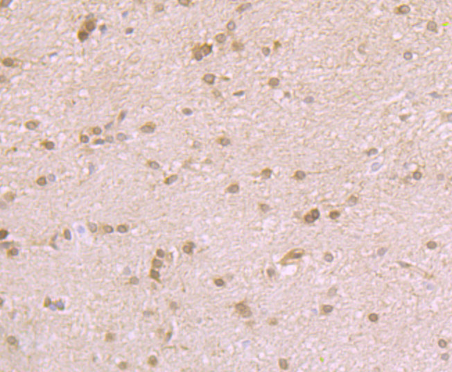
Fig5: Immunohistochemical analysis of paraffin-embedded rat brain tissue using anti-Nesprin 1 antibody. Counter stained with hematoxylin.Application
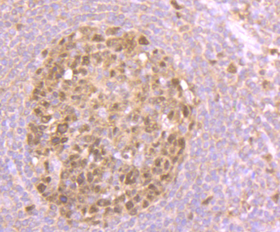
Fig6: Immunohistochemical analysis of paraffin-embedded human tonsil tissue using anti-Nesprin 1 antibody. Counter stained with hematoxylin.Application
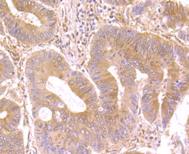
Fig7: Immunohistochemical analysis of paraffin-embedded human colon cancer tissue using anti-Nesprin 1 antibody. Counter stained with hematoxylin.Application
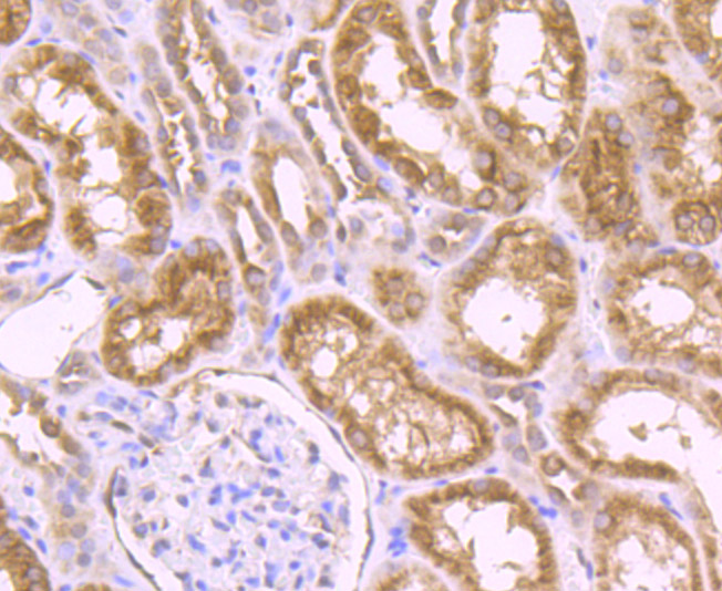
Fig8: Immunohistochemical analysis of paraffin-embedded human kidney tissue using anti-Nesprin 1 antibody. Counter stained with hematoxylin.Application
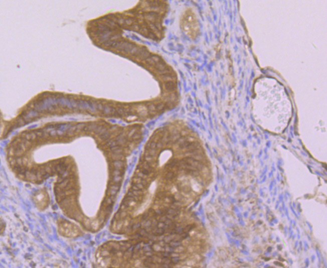
Fig9: Immunohistochemical analysis of paraffin-embedded mouse fallopian tubes tissue using anti-Nesprin 1 antibody. Counter stained with hematoxylin.Application
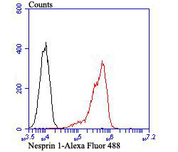
Fig10: Flow cytometric analysis of Daudi cells with Nesprin 1 antibody at 1/100 dilution (red) compared with an unlabelled control (cells without incubation with primary antibody; black).Alexa Fluor 488-conjugated goat anti-rabbit IgG was used as the secondary antibody.| Product Name | Nesprin 1 [JB35-26] |
|---|---|
| Antibody Type | Primary Antibodies |
| Antigen Alias | 8B antibody ARCA1 antibody C6orf98 antibody CPG2 antibody CPG2 full length antibody dJ45H2.2 antibody EDMD4 antibody Enaptin antibody Myne-1 antibody MYNE1 antibody Myocyte nuclear envelope protein 1 antibody Nesp1 antibody Nesprin-1 antibody Nuclear envelope spectrin repeat protein 1 antibody SCAR8 antibody Spectrin repeat containing nuclear envelope 1 antibody Synaptic nuclear envelope protein 1 antibody Synaptic nuclei expressed gene 1 antibody Syne-1 antibody SYNE1 antibody SYNE1_HUMAN antibody SYNE1B antibody |
| Product description | Multi-isomeric modular protein which forms a linking network between organelles and the actin cytoskeleton to maintain the subcellular spatial organization. As a component of the LINC (LInker of Nucleoskeleton and Cytoskeleton) complex involved in the connection between the nuclear lamina and the cytoskeleton. The nucleocytoplasmic interactions established by the LINC complex play an important role in the transmission of mechanical forces across the nuclear envelope and in nuclear movement and positioning. May be involved in nucleus-centrosome attachment and nuclear migration in neural progenitors implicating LINC complex association with SUN1/2 and probably association with cytoplasmic dynein-dynactin motor complexes; SYNE1 and SYNE2 may act redundantly. Required for centrosome migration to the apical cell surface during early ciliogenesis. May be involved in nuclear remodeling during sperm head formation in spermatogenenis; a probable SUN3:SYNE1/KASH1 LINC complex may tether spermatid nuclei to posterior cytoskeletal structures such as the manchette. |
| Immunogen | Recombinant protein |
| Clonality | Monoclonal |
|---|---|
| Isotype | IgG |
| Host Species | Recombinant rabbit |
| Tested Applications | WBICCIFIHCFC |
| WB:1:500-1:2,000 ICC:1:200-1:1,000 IHC:1:50-1:200 FC:1:50-1:100 |
|
| Species Reactivity | HumanMouseRat |
| Concentration | 1mg/ml |
| Alternative Names | 8B antibody ARCA1 antibody C6orf98 antibody CPG2 antibody CPG2 full length antibody dJ45H2.2 antibody EDMD4 antibody Enaptin antibody Myne-1 antibody MYNE1 antibody Myocyte nuclear envelope protein 1 antibody Nesp1 antibody Nesprin-1 antibody Nuclear envelope spectrin repeat protein 1 antibody SCAR8 antibody Spectrin repeat containing nuclear envelope 1 antibody Synaptic nuclear envelope protein 1 antibody Synaptic nuclei expressed gene 1 antibody Syne-1 antibody SYNE1 antibody SYNE1_HUMAN antibody SYNE1B antibody |
|---|---|
| Molecular Weight(MW) | 112 kDa |
| Cellular Localization | Cytoskeleton. Nucleus. |
Application
Fig1: Western blot analysis of Nesprin 1 on different lysates using anti-Nesprin 1 antibody at 1/500 dilution. Positive control: Lane 1: A549 Lane 2: Mouse spleen tissue
Application
Fig2: ICC staining Nesprin 1 in A549 cells (green). The nuclear counter stain is DAPI (blue). Cells were fixed in paraformaldehyde, permeabilised with 0.25% Triton X100/PBS.
Application
Fig3: ICC staining Nesprin 1 in C2C12 cells (green). The nuclear counter stain is DAPI (blue). Cells were fixed in paraformaldehyde, permeabilised with 0.25% Triton X100/PBS.
Application
Fig4: ICC staining Nesprin 1 in HUVEC cells (green). The nuclear counter stain is DAPI (blue). Cells were fixed in paraformaldehyde, permeabilised with 0.25% Triton X100/PBS.
Application
Fig5: Immunohistochemical analysis of paraffin-embedded rat brain tissue using anti-Nesprin 1 antibody. Counter stained with hematoxylin.
Application
Fig6: Immunohistochemical analysis of paraffin-embedded human tonsil tissue using anti-Nesprin 1 antibody. Counter stained with hematoxylin.
Application
Fig7: Immunohistochemical analysis of paraffin-embedded human colon cancer tissue using anti-Nesprin 1 antibody. Counter stained with hematoxylin.
Application
Fig8: Immunohistochemical analysis of paraffin-embedded human kidney tissue using anti-Nesprin 1 antibody. Counter stained with hematoxylin.
Application
Fig9: Immunohistochemical analysis of paraffin-embedded mouse fallopian tubes tissue using anti-Nesprin 1 antibody. Counter stained with hematoxylin.
Application
Fig10: Flow cytometric analysis of Daudi cells with Nesprin 1 antibody at 1/100 dilution (red) compared with an unlabelled control (cells without incubation with primary antibody; black).Alexa Fluor 488-conjugated goat anti-rabbit IgG was used as the secondary antibody.| Positive Control | A549, mouse spleen tissue lysate, C2C12, HUVEC, rat brain tissue, human tonsil tissue, human colon cancer tissue, human kidney tissue, mouse fallopian tubes tissue, Daudi. |
|---|---|
| Application Notes | WB:1:500-1:2,000 ICC:1:200-1:1,000 IHC:1:50-1:200 FC:1:50-1:100 |
| Form | Liquid |
|---|---|
| Storage Instructions | Store at +4℃ after thawing. Aliquot store at -20℃ or -80℃. Avoid repeated freeze / thaw cycles. |
| Storage Buffer | 1*TBS (pH7.4), 1%BSA, 40%Glycerol. Preservative: 0.05% Sodium Azide. |


