Application
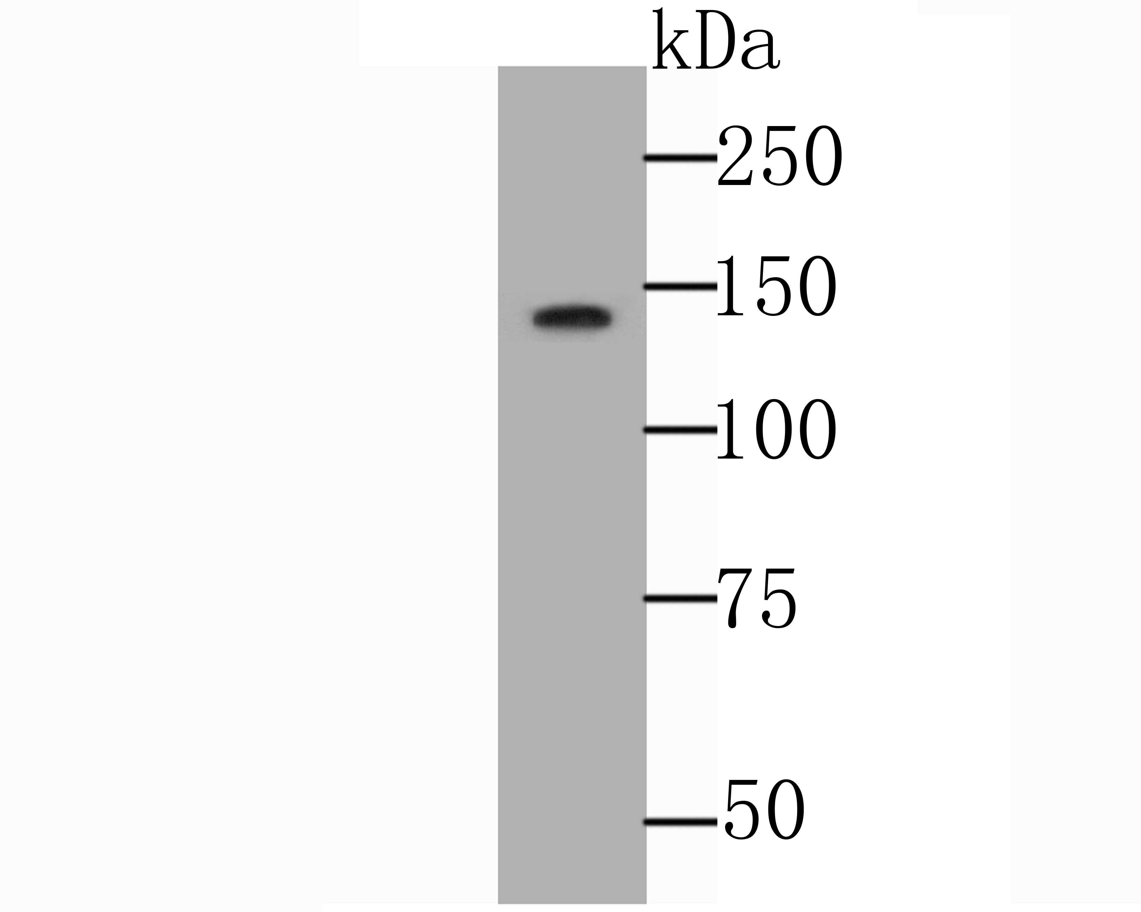
Fig1: Western blot analysis of NRCAM on SH-SY5Y cell lysate using anti-NRCAM antibody at 1/1,000 dilution.Application
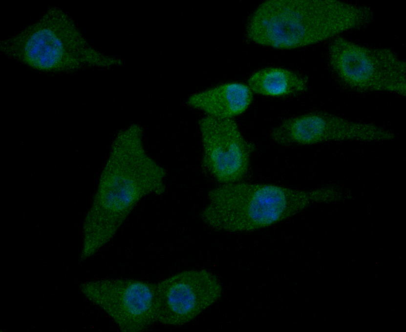
Fig2: ICC staining NRCAM in A549 cells (green). The nuclear counter stain is DAPI (blue). Cells were fixed in paraformaldehyde, permeabilised with 0.25% Triton X100/PBS.Application
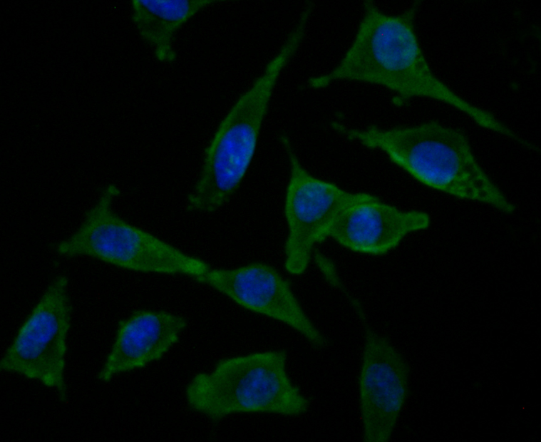
Fig3: ICC staining NRCAM in SH-SY5Y cells (green). The nuclear counter stain is DAPI (blue). Cells were fixed in paraformaldehyde, permeabilised with 0.25% Triton X100/PBS.Application
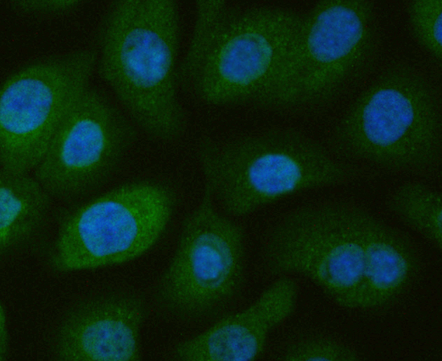
Fig4: ICC staining NRCAM in SKOV-3 cells (green). The nuclear counter stain is DAPI (blue). Cells were fixed in paraformaldehyde, permeabilised with 0.25% Triton X100/PBS.Application
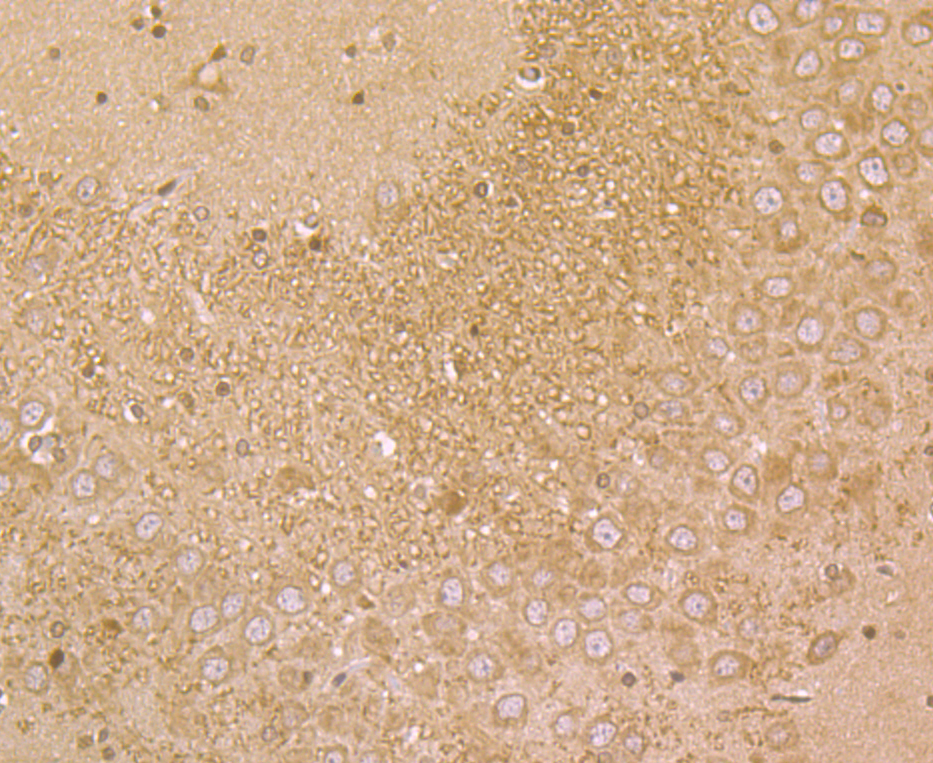
Fig5: Immunohistochemical analysis of paraffin-embedded rat brain tissue using anti-NRCAM antibody. Counter stained with hematoxylin.Application
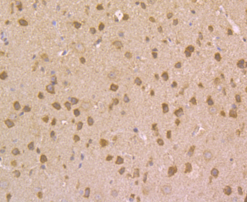
Fig6: Immunohistochemical analysis of paraffin-embedded mouse brain tissue using anti-NRCAM antibody. Counter stained with hematoxylin.| Product Name | NrCAM |
|---|---|
| Antibody Type | Primary Antibodies |
| Antigen Alias | Bravo antibody hBravo antibody Neuronal cell adhesion molecule antibody Neuronal surface protein Bravo antibody Ng CAM related antibody Ng-CAM-related antibody NgCAM related cell adhesion molecule antibody NgCAM-related cell adhesion molecule antibody Nr CAM antibody Nr-CAM antibody Nrcam antibody NRCAM_HUMAN antibody |
| Product description | Cell adhesion protein that is required for normal responses to cell-cell contacts in brain and in the peripheral nervous system. Plays a role in neurite outgrowth in response to contactin binding. Plays a role in mediating cell-cell contacts between Schwann cells and axons. Plays a role in the formation and maintenance of the nodes of Ranvier on myelinated axons. Nodes of Ranvier contain clustered sodium channels that are crucial for the saltatory propagation of action potentials along myelinated axons. During development, nodes of Ranvier are formed by the fusion of two heminodes. Required for normal clustering of sodium channels at heminodes; not required for the formation of mature nodes with normal sodium channel clusters. Required, together with GLDN, for maintaining NFASC and sodium channel clusters at mature nodes of Ranvier. |
| Immunogen | Recombinant protein within human NRCAM aa 700-900. |
| Clonality | Polyclonal |
|---|---|
| Isotype | IgG |
| Host Species | Rabbit |
| Tested Applications | WBICCIHC |
| WB:1:500-1:1,000 ICC:1:50-1:200 IHC:1:50 -1:200 |
|
| Species Reactivity | HumanMouseRat |
| Concentration | 1mg/ml |
| Alternative Names | Bravo antibody hBravo antibody Neuronal cell adhesion molecule antibody Neuronal surface protein Bravo antibody Ng CAM related antibody Ng-CAM-related antibody NgCAM related cell adhesion molecule antibody NgCAM-related cell adhesion molecule antibody Nr CAM antibody Nr-CAM antibody Nrcam antibody NRCAM_HUMAN antibody |
|---|---|
| Molecular Weight(MW) | 144 kDa |
| Cellular Localization | Cell membrane. Secreted. |
Application
Fig1: Western blot analysis of NRCAM on SH-SY5Y cell lysate using anti-NRCAM antibody at 1/1,000 dilution.
Application
Fig2: ICC staining NRCAM in A549 cells (green). The nuclear counter stain is DAPI (blue). Cells were fixed in paraformaldehyde, permeabilised with 0.25% Triton X100/PBS.
Application
Fig3: ICC staining NRCAM in SH-SY5Y cells (green). The nuclear counter stain is DAPI (blue). Cells were fixed in paraformaldehyde, permeabilised with 0.25% Triton X100/PBS.
Application
Fig4: ICC staining NRCAM in SKOV-3 cells (green). The nuclear counter stain is DAPI (blue). Cells were fixed in paraformaldehyde, permeabilised with 0.25% Triton X100/PBS.
Application
Fig5: Immunohistochemical analysis of paraffin-embedded rat brain tissue using anti-NRCAM antibody. Counter stained with hematoxylin.
Application
Fig6: Immunohistochemical analysis of paraffin-embedded mouse brain tissue using anti-NRCAM antibody. Counter stained with hematoxylin.| Positive Control | SH-SY5Y, A549, SKOV-3, rat brain tissue, mouse brain tissue. |
|---|---|
| Application Notes | WB:1:500-1:1,000 ICC:1:50-1:200 IHC:1:50 -1:200 |
| Form | Liquid |
|---|---|
| Storage Instructions | Store at +4℃ after thawing. Aliquot store at -20℃. Avoid repeated freeze / thaw cycles. |
| Storage Buffer | 1*TBS (pH7.4), 0.5%BSA, 50%Glycerol. Preservative: 0.05% Sodium Azide. |


