Application
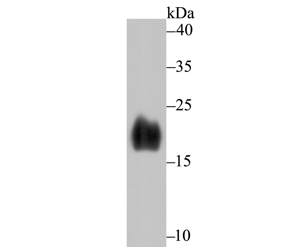
Fig1: Western blot analysis of Alpha Synuclein on human fetal brain tissue lysate using anti-Alpha Synuclein antibody at 1/500 dilution.Application
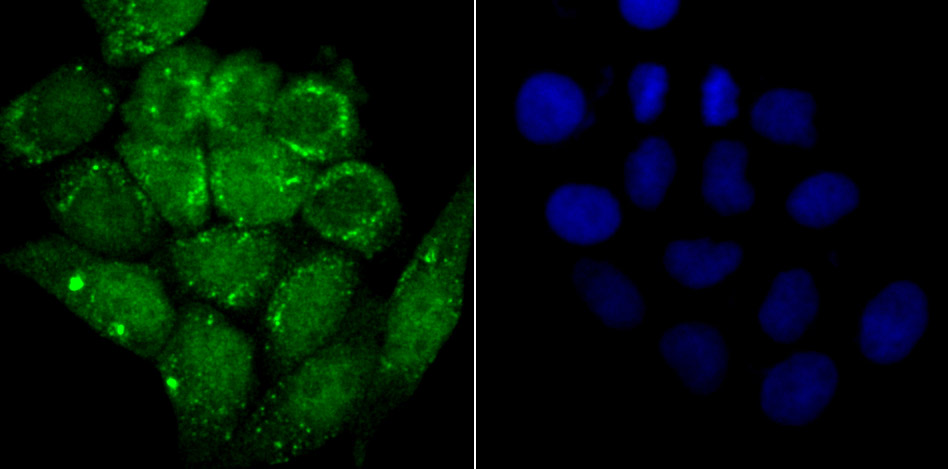
Fig2: ICC staining Alpha Synuclein in Hela cells (green). The nuclear counter stain is DAPI (blue). Cells were fixed in paraformaldehyde, permeabilised with 0.25% Triton X100/PBS.Application
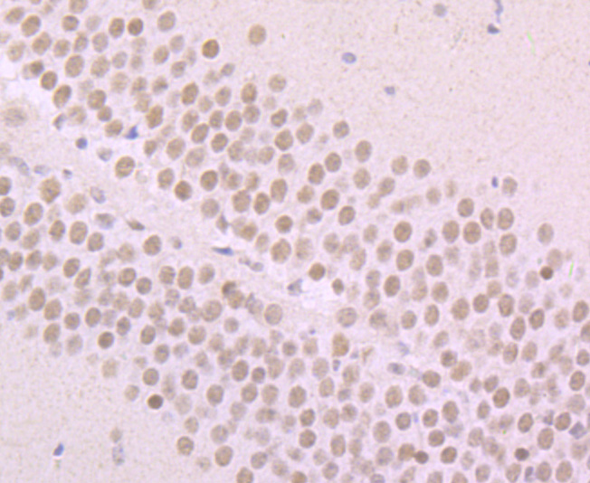
Fig3: Immunohistochemical analysis of paraffin-embedded mouse brain tissue using anti-Alpha Synuclein antibody. Counter stained with hematoxylin.Application
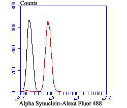
Fig4: Flow cytometric analysis of SH-SY-5Y cells with Alpha Synuclein antibody at 1/100 dilution (red) compared with an unlabelled control (cells without incubation with primary antibody; black). Alexa Fluor 488-conjugated goat anti-rabbit IgG was used as the secondary antibody.| Product Name | Alpha Synuclein [JB42-33] |
|---|---|
| Antibody Type | Primary Antibodies |
| Antigen Alias | Alpha synuclein antibody Alpha-synuclein antibody Alpha-synuclein, isoform NACP140 antibody alphaSYN antibody MGC105443 antibody MGC110988 antibody MGC127560 antibody MGC64356 antibody NACP antibody Non A beta component of AD amyloid antibody Non A4 component of amyloid antibody Non A4 component of amyloid precursor antibody Non-A beta component of AD amyloid antibody Non-A-beta component of alzheimers disease amyloid , precursor of antibody Non-A4 component of amyloid precursor antibody Non-A4 component of amyloid, precursor of antibody OTTHUMP00000218549 antibody OTTHUMP00000218551 antibody OTTHUMP00000218552 antibody OTTHUMP00000218553 antibody OTTHUMP00000218554 antibody PARK 1 antibody PARK 4 antibody PARK1 antibody PARK4 antibody Parkinson disease (autosomal dominant, Lewy body) 4 antibody Parkinson disease familial 1 antibody SNCA antibody Snca synuclein, alpha (non A4 component of amyloid precursor) antibody SYN antibody Synuclein alpha antibody Synuclein alpha 140 antibody Synuclein, alpha (non A4 component of amyloid precursor) antibody SYUA_HUMAN antibody |
| Product description | The synuclein family members, including α-synuclein (also designated NACP for non-β-Amyloid component) and β-synuclein, are predominantly expressed in the brain and are speculated to be involved in synaptic regulation and neuronal plasticity. α-synuclein is localized to neuronal cell bodies and synapses. α-synuclein was first identified as a component of Alzheimer's disease amyloid plaques. Abnormal platelet function in Alzheimer's disease has been demonstrated. During megakaryocytic differentiation α-synuclein has been found to be upregulated, while β-synuclein is downregulated, indicating that coordinate expression of synucleins may be important during hematopoetic cell differentiation. A mutant form of α-synuclein has been found in patients with early onset Parkinson's disease. |
| Immunogen | Recombinant protein |
| Clonality | Monoclonal |
|---|---|
| Isotype | IgG |
| Host Species | Recombinant rabbit |
| Tested Applications | WBICCIFIHCIPFC |
| WB:1:500-1:2,000 ICC:1:50-1:200 IHC:1:50-1:200 FC:1:50-1:100 |
|
| Species Reactivity | HumanMouse |
| Concentration | 1mg/ml |
| Alternative Names | Alpha synuclein antibody
Alpha-synuclein antibody
Alpha-synuclein isoform NACP140 antibody alphaSYN antibody MGC105443 antibody MGC110988 antibody MGC127560 antibody MGC64356 antibody NACP antibody Non A beta component of AD amyloid antibody Non A4 component of amyloid antibody Non A4 component of amyloid precursor antibody Non-A beta component of AD amyloid antibody Non-A-beta component of alzheimers disease amyloid precursor of antibody Non-A4 component of amyloid precursor antibody Non-A4 component of amyloid precursor of antibody OTTHUMP00000218549 antibody OTTHUMP00000218551 antibody OTTHUMP00000218552 antibody OTTHUMP00000218553 antibody OTTHUMP00000218554 antibody PARK 1 antibody PARK 4 antibody PARK1 antibody PARK4 antibody Parkinson disease (autosomal dominant Lewy body) 4 antibody Parkinson disease familial 1 antibody SNCA antibody Snca synuclein alpha (non A4 component of amyloid precursor) antibody SYN antibody Synuclein alpha antibody Synuclein alpha 140 antibody Synuclein alpha (non A4 component of amyloid precursor) antibody SYUA_HUMAN antibody |
|---|---|
| Molecular Weight(MW) | 16 kDa |
| Cellular Localization | Nucleus. Cytoplasm. Secreted. Membrane. |
Application
Fig1: Western blot analysis of Alpha Synuclein on human fetal brain tissue lysate using anti-Alpha Synuclein antibody at 1/500 dilution.
Application
Fig2: ICC staining Alpha Synuclein in Hela cells (green). The nuclear counter stain is DAPI (blue). Cells were fixed in paraformaldehyde, permeabilised with 0.25% Triton X100/PBS.
Application
Fig3: Immunohistochemical analysis of paraffin-embedded mouse brain tissue using anti-Alpha Synuclein antibody. Counter stained with hematoxylin.
Application
Fig4: Flow cytometric analysis of SH-SY-5Y cells with Alpha Synuclein antibody at 1/100 dilution (red) compared with an unlabelled control (cells without incubation with primary antibody; black). Alexa Fluor 488-conjugated goat anti-rabbit IgG was used as the secondary antibody.| Positive Control | Hela, SH-SY-5Y, human fetal brain tissue lysate, mouse brain tissue. |
|---|---|
| Application Notes | WB:1:500-1:2,000 ICC:1:50-1:200 IHC:1:50-1:200 FC:1:50-1:100 |
| Form | Liquid |
|---|---|
| Storage Instructions | Store at +4℃ after thawing. Aliquot store at -20℃ or -80℃. Avoid repeated freeze / thaw cycles. |
| Storage Buffer | 1*TBS (pH7.4), 1%BSA, 40%Glycerol. Preservative: 0.05% Sodium Azide. |


