Application
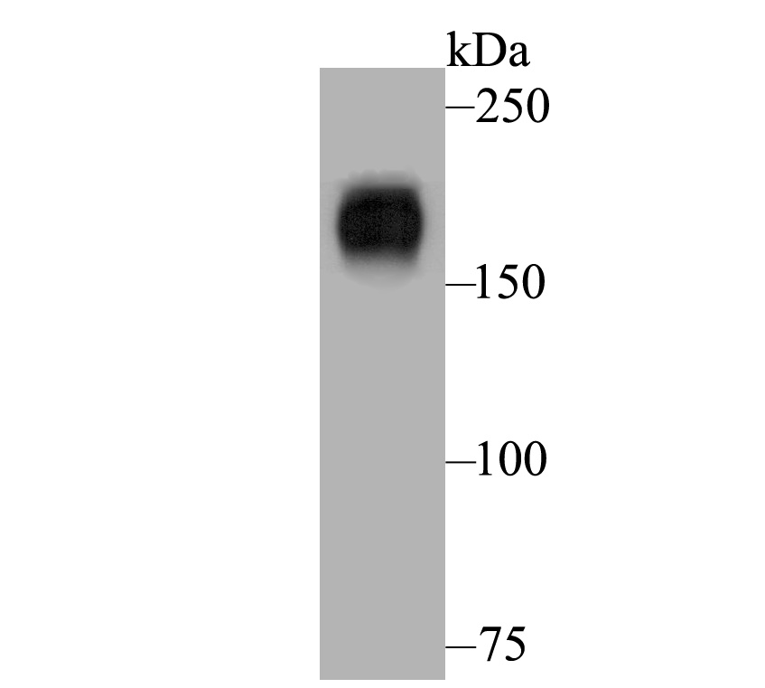
Fig1: Western blot analysis of Nephrin on human kidney lysates. Proteins were transferred to a PVDF membrane and blocked with 5% BSA in PBS for 1 hour at room temperature. The primary antibody was used at a 1:500 dilution in 5% BSA at room temperature for 2 hours. Goat Anti-Rabbit IgG - HRP Secondary Antibody (HA1001) at 1:5,000 dilution was used for 1 hour at room temperature.Application
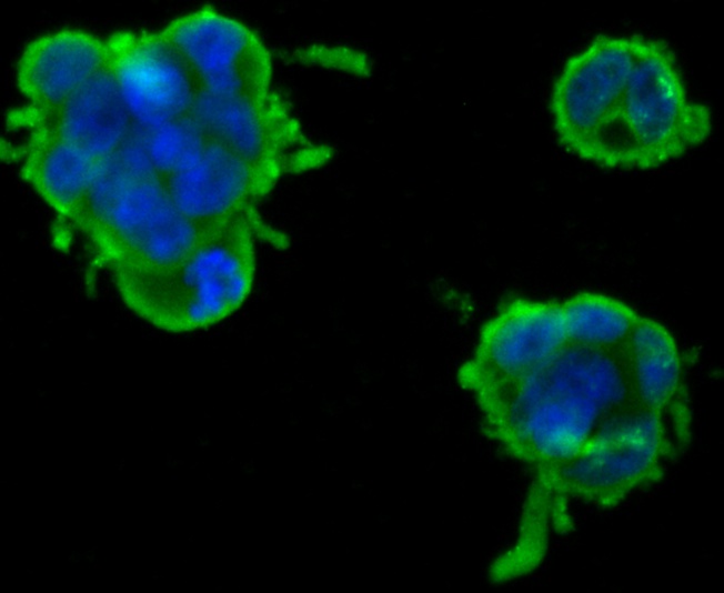
Fig2: ICC staining Nephrin in 293T cells (green). Formalin fixed cells were permeabilized with 0.1% Triton X-100 in TBS for 10 minutes at room temperature and blocked with 1% Blocker BSA for 15 minutes at room temperature. Cells were probed with Nephrin polyclonal antibody at a dilution of 1:100 for at least 1 hour at room temperature, washed with PBS. Alexa Fluorc™ 488 Goat anti-Rabbit IgG was used as the secondary antibody at 1/100 dilution. The nuclear counter stain is DAPI (blue).Application
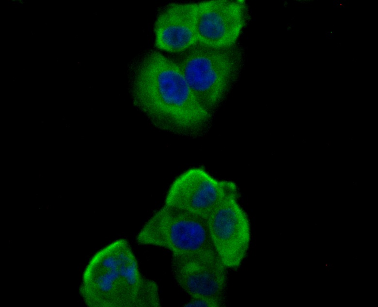
Fig3: ICC staining Nephrin in PANC-1 cells (green). Formalin fixed cells were permeabilized with 0.1% Triton X-100 in TBS for 10 minutes at room temperature and blocked with 1% Blocker BSA for 15 minutes at room temperature. Cells were probed with Nephrin polyclonal antibody at a dilution of 1:100 for at least 1 hour at room temperature, washed with PBS. Alexa Fluorc™ 488 Goat anti-Rabbit IgG was used as the secondary antibody at 1/100 dilution. The nuclear counter stain is DAPI (blue).Application
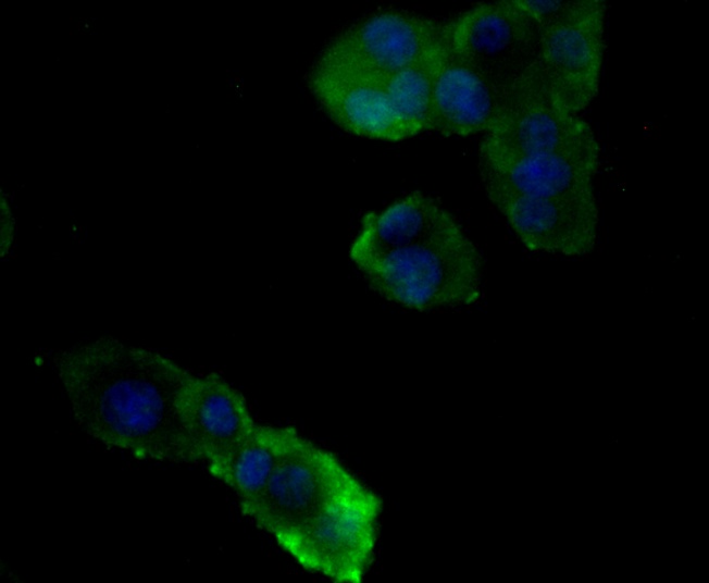
Fig4: ICC staining Nephrin in PC-12 cells (green). Formalin fixed cells were permeabilized with 0.1% Triton X-100 in TBS for 10 minutes at room temperature and blocked with 1% Blocker BSA for 15 minutes at room temperature. Cells were probed with Nephrin polyclonal antibody at a dilution of 1:100 for at least 1 hour at room temperature, washed with PBS. Alexa Fluorc™ 488 Goat anti-Rabbit IgG was used as the secondary antibody at 1/100 dilution. The nuclear counter stain is DAPI (blue).| Product Name | Nephrin |
|---|---|
| Antibody Type | Primary Antibodies |
| Antigen Alias | CNF antibody Nephrin antibody Nephrosis 1 congenital Finnish type antibody Nephrosis 1, congenital, Finnish type (nephrin) antibody NPHN antibody NPHN_HUMAN antibody NPHS 1 antibody Nphs1 antibody Renal glomerulus specific cell adhesion receptor antibody Renal glomerulus-specific cell adhesion receptor antibody |
| Product description | This gene encodes a member of the immunoglobulin family of cell adhesion molecules that functions in the glomerular filtration barrier in the kidney. The renal filtration barrier consists of fenestrated endothelial cells, the glomerular basement membrane, and the podocytes of epithelial cells. The gene is primarily expressed in renal tissues, and the protein is a type-1 transmembrane protein found at the slit diaphragm of glomerular podocytes. The slit diaphragm is thought to function as an ultrafilter to exclude albumin and other plasma macromolecules in the formation of urine. Mutations in this gene result in Finnish-type congenital nephrosis 1, characterized by severe proteinuria and loss of the slit diaphragm and foot processes. A defect in the gene for nephrin, NPHS1, is associated with congenital nephrotic syndrome of the Finnish type and causes massive amounts of protein to be leaked into the urine, or proteinuria. Nephrin is also required for cardiovascular development. Plays a role in skeletal muscle formation through regulation of myoblast fusion. |
| Immunogen | Recombinant protein within human Nephrin aa 1000-1250. |
| Clonality | Polyclonal |
|---|---|
| Isotype | IgG |
| Host Species | Rabbit |
| Tested Applications | WBICC |
| WB:1:500-1:2,000: ICC:1:50-1:200: |
|
| Species Reactivity | HumanRat |
| Concentration | 1mg/ml |
| Alternative Names | CNF antibody
Nephrin antibody
Nephrosis 1 congenital Finnish type antibody
Nephrosis 1 congenital Finnish type (nephrin) antibody NPHN antibody NPHN_HUMAN antibody NPHS 1 antibody Nphs1 antibody Renal glomerulus specific cell adhesion receptor antibody Renal glomerulus-specific cell adhesion receptor antibody |
|---|---|
| Molecular Weight(MW) | 160 kDa, predicted band size 136 kDa |
| Cellular Localization | Cell membrane, Membrane. |
Application
Fig1: Western blot analysis of Nephrin on human kidney lysates. Proteins were transferred to a PVDF membrane and blocked with 5% BSA in PBS for 1 hour at room temperature. The primary antibody was used at a 1:500 dilution in 5% BSA at room temperature for 2 hours. Goat Anti-Rabbit IgG - HRP Secondary Antibody (HA1001) at 1:5,000 dilution was used for 1 hour at room temperature.
Application
Fig2: ICC staining Nephrin in 293T cells (green). Formalin fixed cells were permeabilized with 0.1% Triton X-100 in TBS for 10 minutes at room temperature and blocked with 1% Blocker BSA for 15 minutes at room temperature. Cells were probed with Nephrin polyclonal antibody at a dilution of 1:100 for at least 1 hour at room temperature, washed with PBS. Alexa Fluorc™ 488 Goat anti-Rabbit IgG was used as the secondary antibody at 1/100 dilution. The nuclear counter stain is DAPI (blue).
Application
Fig3: ICC staining Nephrin in PANC-1 cells (green). Formalin fixed cells were permeabilized with 0.1% Triton X-100 in TBS for 10 minutes at room temperature and blocked with 1% Blocker BSA for 15 minutes at room temperature. Cells were probed with Nephrin polyclonal antibody at a dilution of 1:100 for at least 1 hour at room temperature, washed with PBS. Alexa Fluorc™ 488 Goat anti-Rabbit IgG was used as the secondary antibody at 1/100 dilution. The nuclear counter stain is DAPI (blue).
Application
Fig4: ICC staining Nephrin in PC-12 cells (green). Formalin fixed cells were permeabilized with 0.1% Triton X-100 in TBS for 10 minutes at room temperature and blocked with 1% Blocker BSA for 15 minutes at room temperature. Cells were probed with Nephrin polyclonal antibody at a dilution of 1:100 for at least 1 hour at room temperature, washed with PBS. Alexa Fluorc™ 488 Goat anti-Rabbit IgG was used as the secondary antibody at 1/100 dilution. The nuclear counter stain is DAPI (blue).| Positive Control | Human kidney tissue, 293T, PANC-1, PC-12. |
|---|---|
| Application Notes | WB:1:500-1:2,000: ICC:1:50-1:200: |
| Form | Liquid |
|---|---|
| Storage Instructions | Store at +4℃ after thawing. Aliquot store at -20℃. Avoid repeated freeze / thaw cycles. |
| Storage Buffer | 1*TBS (pH7.4), 1%BSA, 50%Glycerol. Preservative: 0.05% Sodium Azide. |


