Application
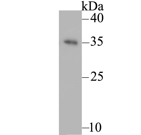
Fig1: Western blot analysis of GTSF1 on zebrafish lysate. Proteins were transferred to a PVDF membrane and blocked with 5% BSA in PBS for 1 hour at room temperature. The primary antibody was used at a 1:200 dilution in 5% BSA at room temperature for 2 hours. Goat Anti-Rabbit IgG - HRP Secondary Antibody (HA1001) at 1:5,000 dilution was used for 1 hour at room temperature.Application
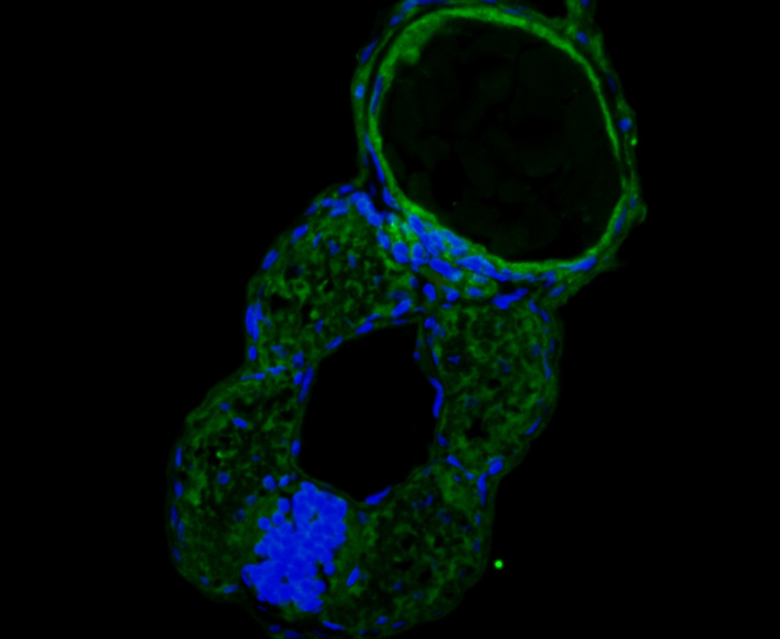
Fig2: Immunofluorescence staining of paraffin-embedded Zebrafish using anti-GTSF1 rabbit polyclonal antibody.The section was pre-treated using heat mediated antigen retrieval with Tris-EDTA buffer (pH 9.0) for 20 minutes. The tissues were blocked in 10% negative goat serum for 1 hour at room temperature, washed with PBS, and then probed with the antibody (ER1803-68) at 1/50 dilution for 10 hours at 4℃ and detected using Alexa Fluor™ 488 conjugate-Goat anti-Rabbit IgG (H+L) Secondary Antibody at a dilution of 1:500 for 1 hour at room temperature.Application
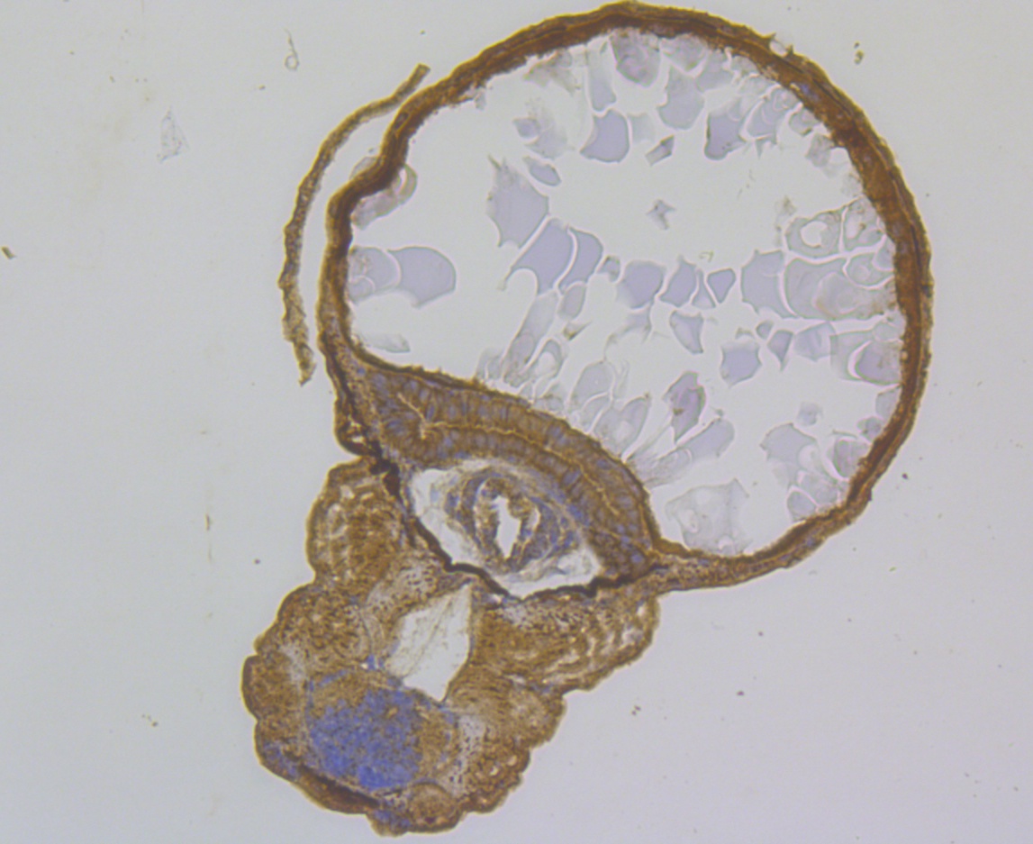
Fig3: Immunohistochemical analysis of paraffin-embedded Zebrafish tissue using anti-GTSF1 antibody. The section was pre-treated using heat mediated antigen retrieval with Tris-EDTA buffer (pH 8.0-8.4) for 20 minutes.The tissues were blocked in 5% BSA for 30 minutes at room temperature, washed with ddH2O and PBS, and then probed with the antibody (ER1803-68) at 1/50 dilution, for 30 minutes at room temperature and detected using an HRP conjugated compact polymer system. DAB was used as the chrogen. Counter stained with hematoxylin and mounted with DPX.| Product Name | GTSF1 |
|---|---|
| Antibody Type | Primary Antibodies |
| Antigen Alias | Cue110 antibody Fam112b antibody Family with sequence similarity 112 member B antibody FLJ32942 antibody Gametocyte-specific factor 1 antibody GTSF1 antibody GTSF1_HUMAN antibody Protein FAM112B antibody |
| Product description | Gametocyte-specific factor 1 (Gtsf1), also known as the computationally obtained undifferentiated and/or embryonic stem cell-specific 110 gene (Cue110). Gtsf1 encodes a 167-amino acid protein that is a member of the Uncharacterized Protein Family 0224. DmGTSF1 is essential for P-element-induced wimpy testis (PIWI)-interacting RNA (piRNA)-mediated transcriptional repression and histone H3K9me3-mediated repression of transposons and their neighboring genes in the ovary. DmGTSF1 interacts with Piwi via its C-terminal tail and the two CHHC zinc-finger motifs within the DmGTSF1 protein are required for its activity. The Gtsf1 gene is essential for spermatogenesis and has been revealed to function in transposon suppression in the mouse testes. GTSF1 transcript expression has been observed to be up-regulated in certain leukemias and in lymphomas. |
| Immunogen | Recombinant protein within zebrafish GTSF1 aa 20-170. |
| Clonality | Polyclonal |
|---|---|
| Isotype | IgG |
| Host Species | Rabbit |
| Tested Applications | WBIFIHC |
| WB:1:200: IF:1:50-1:200: IHC:1:50-1:200: |
|
| Species Reactivity | Zebra Fish |
| Concentration | 1mg/ml |
| Alternative Names | Cue110 antibody Fam112b antibody Family with sequence similarity 112 member B antibody FLJ32942 antibody Gametocyte-specific factor 1 antibody GTSF1 antibody GTSF1_HUMAN antibody Protein FAM112B antibody |
|---|---|
| Molecular Weight(MW) | 35 kDa, predicted band size 21 kDa |
| Cellular Localization | Cytoplasm. |
| SwissProt ID | K4FBI1 |
|---|
Application
Fig1: Western blot analysis of GTSF1 on zebrafish lysate. Proteins were transferred to a PVDF membrane and blocked with 5% BSA in PBS for 1 hour at room temperature. The primary antibody was used at a 1:200 dilution in 5% BSA at room temperature for 2 hours. Goat Anti-Rabbit IgG - HRP Secondary Antibody (HA1001) at 1:5,000 dilution was used for 1 hour at room temperature.
Application
Fig2: Immunofluorescence staining of paraffin-embedded Zebrafish using anti-GTSF1 rabbit polyclonal antibody.The section was pre-treated using heat mediated antigen retrieval with Tris-EDTA buffer (pH 9.0) for 20 minutes. The tissues were blocked in 10% negative goat serum for 1 hour at room temperature, washed with PBS, and then probed with the antibody (ER1803-68) at 1/50 dilution for 10 hours at 4℃ and detected using Alexa Fluor™ 488 conjugate-Goat anti-Rabbit IgG (H+L) Secondary Antibody at a dilution of 1:500 for 1 hour at room temperature.
Application
Fig3: Immunohistochemical analysis of paraffin-embedded Zebrafish tissue using anti-GTSF1 antibody. The section was pre-treated using heat mediated antigen retrieval with Tris-EDTA buffer (pH 8.0-8.4) for 20 minutes.The tissues were blocked in 5% BSA for 30 minutes at room temperature, washed with ddH2O and PBS, and then probed with the antibody (ER1803-68) at 1/50 dilution, for 30 minutes at room temperature and detected using an HRP conjugated compact polymer system. DAB was used as the chrogen. Counter stained with hematoxylin and mounted with DPX.| Positive Control | Zebrafish. |
|---|---|
| Application Notes | WB:1:200: IF:1:50-1:200: IHC:1:50-1:200: |
| Form | Liquid |
|---|---|
| Storage Instructions | Store at +4℃ after thawing. Aliquot store at -20℃. Avoid repeated freeze / thaw cycles. |
| Storage Buffer | 1*TBS (pH7.4), 1%BSA, 50%Glycerol. Preservative: 0.05% Sodium Azide. |


