Application
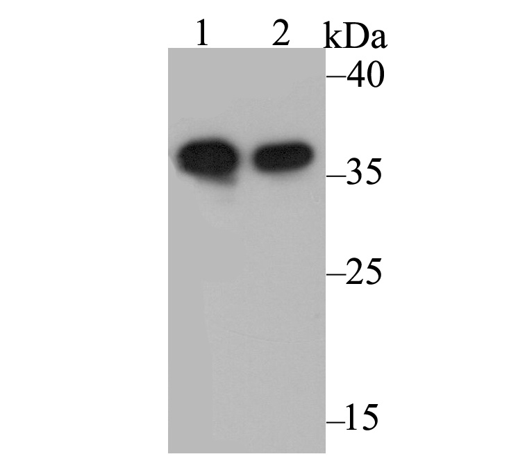
Fig1: Western blot analysis of Annexin A1 on different lysates. Proteins were transferred to a PVDF membrane and blocked with 5% BSA in PBS for 1 hour at room temperature. The primary antibody was used at a 1:500 dilution in 5% BSA at room temperature for 2 hours. Goat Anti-Rabbit IgG - HRP Secondary Antibody (HA1001) at 1:5,000 dilution was used for 1 hour at room temperature. Positive control: Lane 1: A431 cell lysate Lane 2: Rat uterus tissue lysateApplication
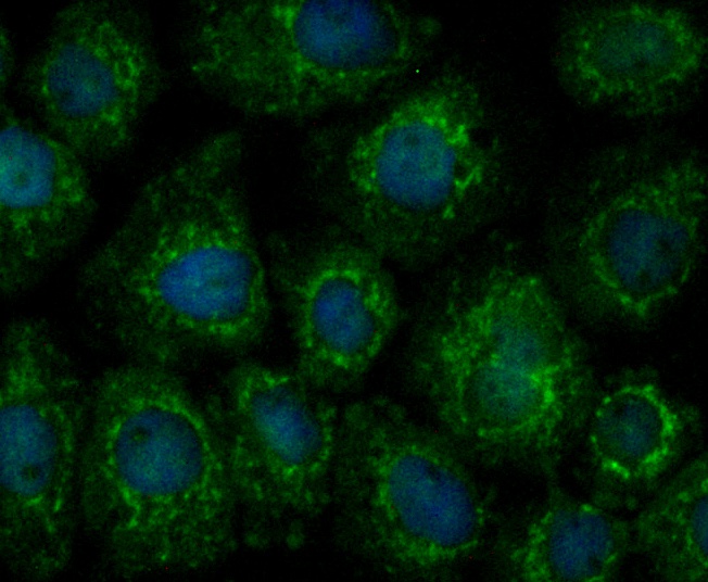
Fig2: ICC staining Annexin A1 in A431 cells (green). Formalin fixed cells were permeabilized with 0.1% Triton X-100 in TBS for 10 minutes at room temperature and blocked with 1% Blocker BSA for 15 minutes at room temperature. Cells were probed with Annexin A1 polyclonal antibody at a dilution of 1:200 for at least 1 hour at room temperature, washed with PBS. Alexa Fluorc™ 488 Goat anti-Rabbit IgG was used as the secondary antibody at 1/100 dilution. The nuclear counter stain is DAPI (blue).Application
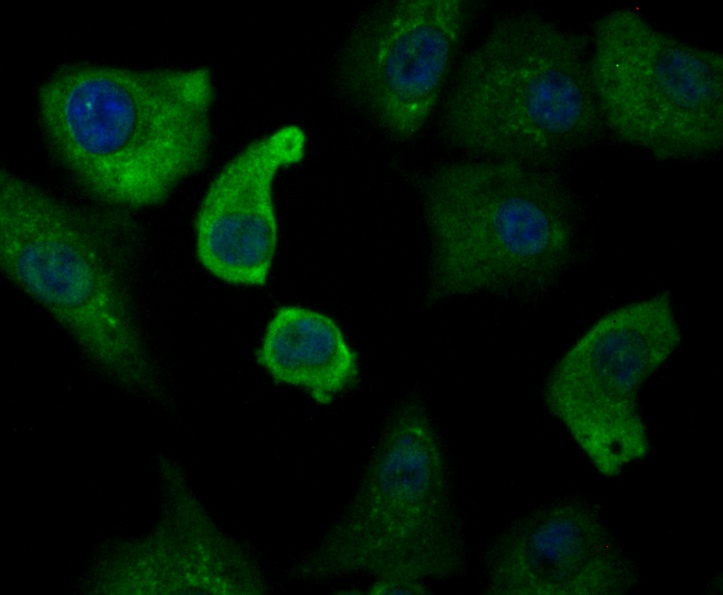
Fig3: ICC staining Annexin A1 in A549 cells (green). Formalin fixed cells were permeabilized with 0.1% Triton X-100 in TBS for 10 minutes at room temperature and blocked with 1% Blocker BSA for 15 minutes at room temperature. Cells were probed with Annexin A1 polyclonal antibody at a dilution of 1:50 for at least 1 hour at room temperature, washed with PBS. Alexa Fluorc™ 488 Goat anti-Rabbit IgG was used as the secondary antibody at 1/100 dilution. The nuclear counter stain is DAPI (blue).Application
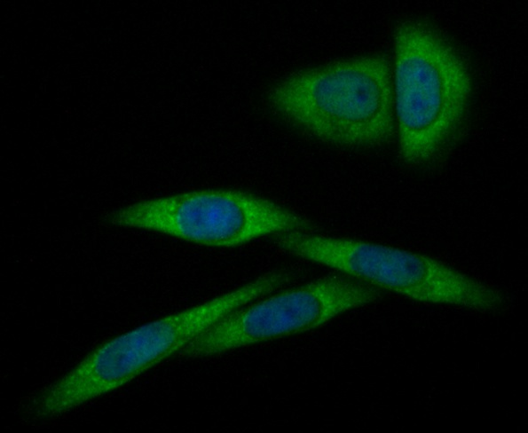
Fig4: ICC staining Annexin A1 in SiHa cells (green). Formalin fixed cells were permeabilized with 0.1% Triton X-100 in TBS for 10 minutes at room temperature and blocked with 1% Blocker BSA for 15 minutes at room temperature. Cells were probed with Annexin A1 polyclonal antibody at a dilution of 1:100 for at least 1 hour at room temperature, washed with PBS. Alexa Fluorc™ 488 Goat anti-Rabbit IgG was used as the secondary antibody at 1/100 dilution. The nuclear counter stain is DAPI (blue).Application
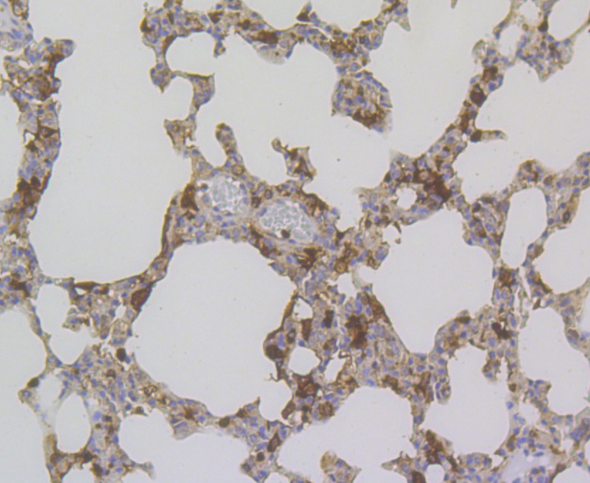
Fig5: Immunohistochemical analysis of paraffin-embedded rat lung tissue using anti-Annexin A1 antibody. The section was pre-treated using heat mediated antigen retrieval with sodium citrate buffer (pH 6.0) for 20 minutes. The tissues were blocked in 5% BSA for 30 minutes at room temperature, washed with ddH2O and PBS, and then probed with the antibody (ER1803-70) at 1/200 dilution, for 30 minutes at room temperature and detected using an HRP conjugated compact polymer system. DAB was used as the chrogen. Counter stained with hematoxylin and mounted with DPX.Application
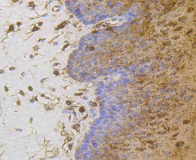
Fig6: Immunohistochemical analysis of paraffin-embedded human esophagus tissue using anti-Annexin A1 antibody. The section was pre-treated using heat mediated antigen retrieval with sodium citrate buffer (pH 6.0) for 20 minutes. The tissues were blocked in 5% BSA for 30 minutes at room temperature, washed with ddH2O and PBS, and then probed with the antibody (ER1803-70) at 1/200 dilution, for 30 minutes at room temperature and detected using an HRP conjugated compact polymer system. DAB was used as the chrogen. Counter stained with hematoxylin and mounted with DPX.Application
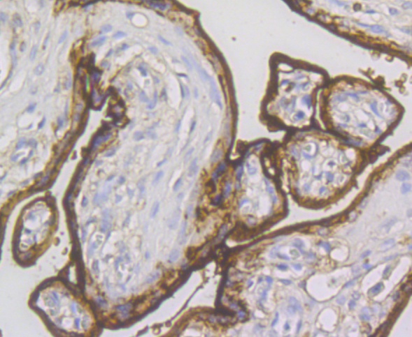
Fig7: Immunohistochemical analysis of paraffin-embedded human placenta tissue using anti-Annexin A1 antibody. The section was pre-treated using heat mediated antigen retrieval with sodium citrate buffer (pH 6.0) for 20 minutes. The tissues were blocked in 5% BSA for 30 minutes at room temperature, washed with ddH2O and PBS, and then probed with the antibody (ER1803-70) at 1/200 dilution, for 30 minutes at room temperature and detected using an HRP conjugated compact polymer system. DAB was used as the chrogen. Counter stained with hematoxylin and mounted with DPX.Application
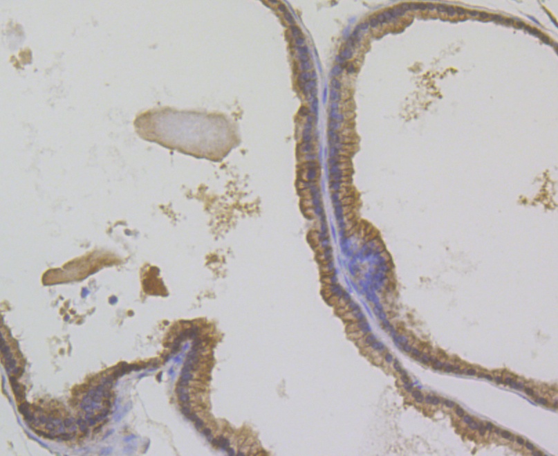
Fig8: Immunohistochemical analysis of paraffin-embedded mouse prostate tissue using anti-Annexin A1 antibody. The section was pre-treated using heat mediated antigen retrieval with sodium citrate buffer (pH 6.0) for 20 minutes. The tissues were blocked in 5% BSA for 30 minutes at room temperature, washed with ddH2O and PBS, and then probed with the antibody (ER1803-70) at 1/200 dilution, for 30 minutes at room temperature and detected using an HRP conjugated compact polymer system. DAB was used as the chrogen. Counter stained with hematoxylin and mounted with DPX.| Product Name | Annexin A1 |
|---|---|
| Antibody Type | Primary Antibodies |
| Antigen Alias | Annexin 1 antibody Annexin A1 antibody Annexin I (lipocortin I) antibody Annexin I antibody Annexin-1 antibody AnnexinA1 antibody AnnexinI antibody ANX 1 antibody ANX A1 antibody ANX1 antibody ANXA 1 antibody ANXA1 antibody ANXA1 protein antibody ANXA1_HUMAN antibody Calpactin 2 antibody Calpactin II antibody Calpactin-2 antibody CalpactinII antibody Chromobindin 9 antibody Chromobindin-9 antibody Chromobindin9 antibody HGNC:533 antibody Lipocortin 1 antibody Lipocortin I antibody Lipocortin1 antibody LipocortinI antibody LPC 1 antibody LPC1 antibody p35 antibody Phospholipase A2 inhibitory protein antibody |
| Product description | Plays important roles in the innate immune response as effector of glucocorticoid-mediated responses and regulator of the inflammatory process. Has anti-inflammatory activity. Plays a role in glucocorticoid-mediated down-regulation of the early phase of the inflammatory response. Promotes resolution of inflammation and wound healing. Functions at least in part by activating the formyl peptide receptors and downstream signaling cascades. Promotes chemotaxis of granulocytes and monocytes via activation of the formyl peptide receptors. Contributes to the adaptive immune response by enhancing signaling cascades that are triggered by T-cell activation, regulates differentiation and proliferation of activated T-cells. Promotes the differentiation of T-cells into Th1 cells and negatively regulates differentiation into Th2 cells. Has no effect on unstimulated T cells. Promotes rearrangement of the actin cytoskeleton, cell polarization and cell migration. Negatively regulates hormone exocytosis via activation of the formyl peptide receptors and reorganization of the actin cytoskeleton. Has high affinity for Ca2+ and can bind up to eight Ca2+ ions. Displays Ca2+-dependent binding to phospholipid membranes. Plays a role in the formation of phagocytic cups and phagosomes. Plays a role in phagocytosis by mediating the Ca2+-dependent interaction between phagosomes and the actin cytoskeleton. |
| Immunogen | Recombinant protein within human Annexin A1 aa 150-310. |
| Clonality | Polyclonal |
|---|---|
| Isotype | IgG |
| Host Species | Rabbit |
| Tested Applications | WBICCIHC |
| WB:1:500-1:2,000 ICC:1:50-1:200: IHC:1:200: |
|
| Species Reactivity | HumanMouseRat |
| Concentration | 1mg/ml |
| Alternative Names | Annexin 1 antibody Annexin A1 antibody Annexin I (lipocortin I) antibody Annexin I antibody Annexin-1 antibody AnnexinA1 antibody AnnexinI antibody ANX 1 antibody ANX A1 antibody ANX1 antibody ANXA 1 antibody ANXA1 antibody ANXA1 protein antibody ANXA1_HUMAN antibody Calpactin 2 antibody Calpactin II antibody Calpactin-2 antibody CalpactinII antibody Chromobindin 9 antibody Chromobindin-9 antibody Chromobindin9 antibody HGNC:533 antibody Lipocortin 1 antibody Lipocortin I antibody Lipocortin1 antibody LipocortinI antibody LPC 1 antibody LPC1 antibody p35 antibody Phospholipase A2 inhibitory protein antibody |
|---|---|
| Molecular Weight(MW) | 39 kDa |
| Cellular Localization | Cell membrane, Cell projection, Cilium, Cytoplasm, Cytoplasmic vesicle, Endosome, Membrane, Nucleus, Secreted. |
Application
Fig1: Western blot analysis of Annexin A1 on different lysates. Proteins were transferred to a PVDF membrane and blocked with 5% BSA in PBS for 1 hour at room temperature. The primary antibody was used at a 1:500 dilution in 5% BSA at room temperature for 2 hours. Goat Anti-Rabbit IgG - HRP Secondary Antibody (HA1001) at 1:5,000 dilution was used for 1 hour at room temperature. Positive control: Lane 1: A431 cell lysate Lane 2: Rat uterus tissue lysate
Application
Fig2: ICC staining Annexin A1 in A431 cells (green). Formalin fixed cells were permeabilized with 0.1% Triton X-100 in TBS for 10 minutes at room temperature and blocked with 1% Blocker BSA for 15 minutes at room temperature. Cells were probed with Annexin A1 polyclonal antibody at a dilution of 1:200 for at least 1 hour at room temperature, washed with PBS. Alexa Fluorc™ 488 Goat anti-Rabbit IgG was used as the secondary antibody at 1/100 dilution. The nuclear counter stain is DAPI (blue).
Application
Fig3: ICC staining Annexin A1 in A549 cells (green). Formalin fixed cells were permeabilized with 0.1% Triton X-100 in TBS for 10 minutes at room temperature and blocked with 1% Blocker BSA for 15 minutes at room temperature. Cells were probed with Annexin A1 polyclonal antibody at a dilution of 1:50 for at least 1 hour at room temperature, washed with PBS. Alexa Fluorc™ 488 Goat anti-Rabbit IgG was used as the secondary antibody at 1/100 dilution. The nuclear counter stain is DAPI (blue).
Application
Fig4: ICC staining Annexin A1 in SiHa cells (green). Formalin fixed cells were permeabilized with 0.1% Triton X-100 in TBS for 10 minutes at room temperature and blocked with 1% Blocker BSA for 15 minutes at room temperature. Cells were probed with Annexin A1 polyclonal antibody at a dilution of 1:100 for at least 1 hour at room temperature, washed with PBS. Alexa Fluorc™ 488 Goat anti-Rabbit IgG was used as the secondary antibody at 1/100 dilution. The nuclear counter stain is DAPI (blue).
Application
Fig5: Immunohistochemical analysis of paraffin-embedded rat lung tissue using anti-Annexin A1 antibody. The section was pre-treated using heat mediated antigen retrieval with sodium citrate buffer (pH 6.0) for 20 minutes. The tissues were blocked in 5% BSA for 30 minutes at room temperature, washed with ddH2O and PBS, and then probed with the antibody (ER1803-70) at 1/200 dilution, for 30 minutes at room temperature and detected using an HRP conjugated compact polymer system. DAB was used as the chrogen. Counter stained with hematoxylin and mounted with DPX.
Application
Fig6: Immunohistochemical analysis of paraffin-embedded human esophagus tissue using anti-Annexin A1 antibody. The section was pre-treated using heat mediated antigen retrieval with sodium citrate buffer (pH 6.0) for 20 minutes. The tissues were blocked in 5% BSA for 30 minutes at room temperature, washed with ddH2O and PBS, and then probed with the antibody (ER1803-70) at 1/200 dilution, for 30 minutes at room temperature and detected using an HRP conjugated compact polymer system. DAB was used as the chrogen. Counter stained with hematoxylin and mounted with DPX.
Application
Fig7: Immunohistochemical analysis of paraffin-embedded human placenta tissue using anti-Annexin A1 antibody. The section was pre-treated using heat mediated antigen retrieval with sodium citrate buffer (pH 6.0) for 20 minutes. The tissues were blocked in 5% BSA for 30 minutes at room temperature, washed with ddH2O and PBS, and then probed with the antibody (ER1803-70) at 1/200 dilution, for 30 minutes at room temperature and detected using an HRP conjugated compact polymer system. DAB was used as the chrogen. Counter stained with hematoxylin and mounted with DPX.
Application
Fig8: Immunohistochemical analysis of paraffin-embedded mouse prostate tissue using anti-Annexin A1 antibody. The section was pre-treated using heat mediated antigen retrieval with sodium citrate buffer (pH 6.0) for 20 minutes. The tissues were blocked in 5% BSA for 30 minutes at room temperature, washed with ddH2O and PBS, and then probed with the antibody (ER1803-70) at 1/200 dilution, for 30 minutes at room temperature and detected using an HRP conjugated compact polymer system. DAB was used as the chrogen. Counter stained with hematoxylin and mounted with DPX.| Positive Control | A431, rat uterus tissue, A549, SiHa, rat lung tissue, human esophagus tissue, human placenta tissue, mouse prostate tissue. |
|---|---|
| Application Notes | WB:1:500-1:2,000 ICC:1:50-1:200: IHC:1:200: |
| Form | Liquid |
|---|---|
| Storage Instructions | Store at +4℃ after thawing. Aliquot store at -20℃. Avoid repeated freeze / thaw cycles. |
| Storage Buffer | 1*TBS (pH7.4), 1%BSA, 50%Glycerol. Preservative: 0.05% Sodium Azide. |


