Application
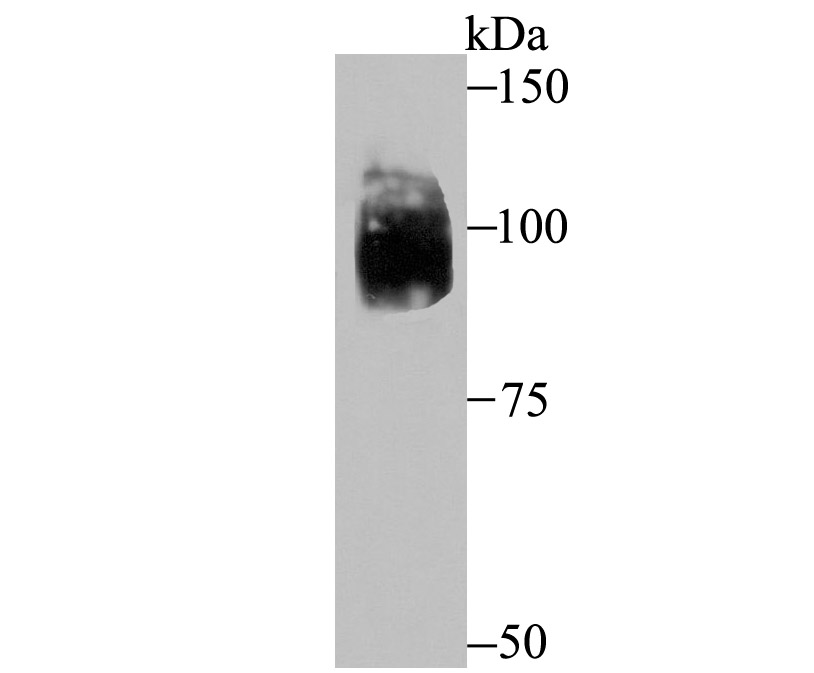
Fig1: Western blot analysis of HIF-2 alpha on SiHa cell lysate using anti-HIF-2 alpha antibody at 1/500 dilution.Application
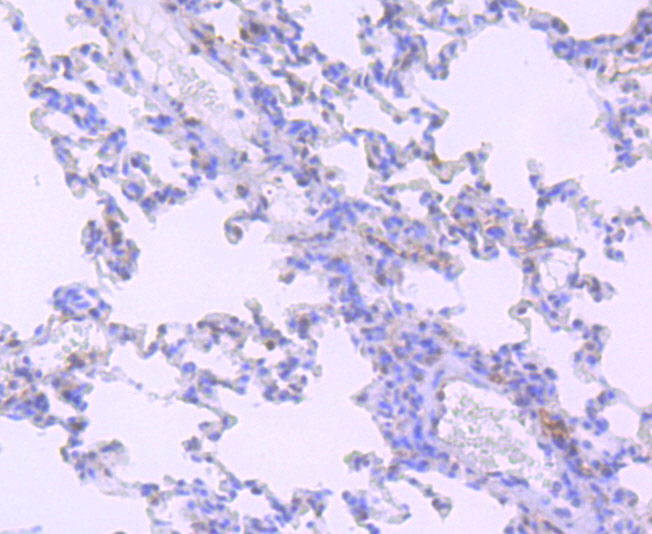
Fig2: Immunohistochemical analysis of paraffin-embedded rat lung tissue using anti-HIF-2 alpha antibody. Counter stained with hematoxylin.Application
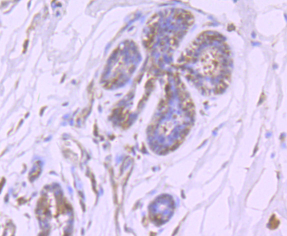
Fig3: Immunohistochemical analysis of paraffin-embedded human breast cancer tissue using anti-HIF-2 alpha antibody. Counter stained with hematoxylin.Application
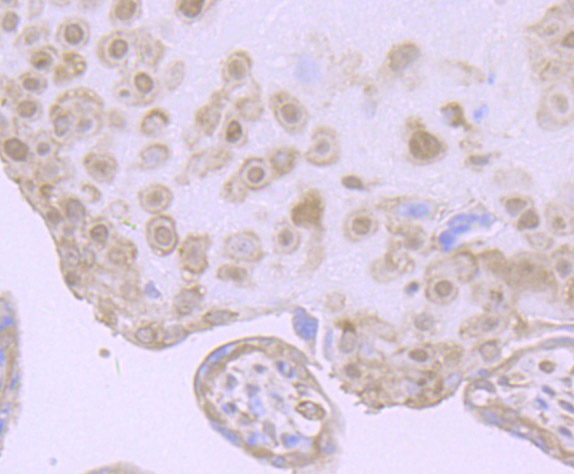
Fig4: Immunohistochemical analysis of paraffin-embedded human placenta tissue using anti-HIF-2 alpha antibody. Counter stained with hematoxylin.Application
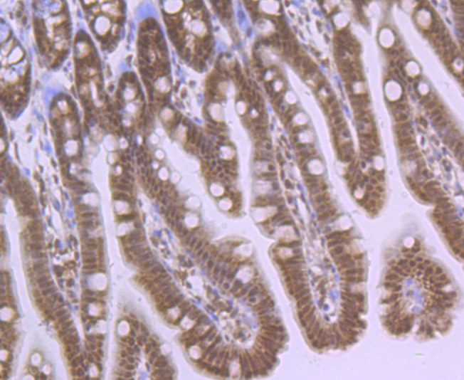
Fig5: Immunohistochemical analysis of paraffin-embedded mouse colon tissue using anti-HIF-2 alpha antibody. Counter stained with hematoxylin.Application
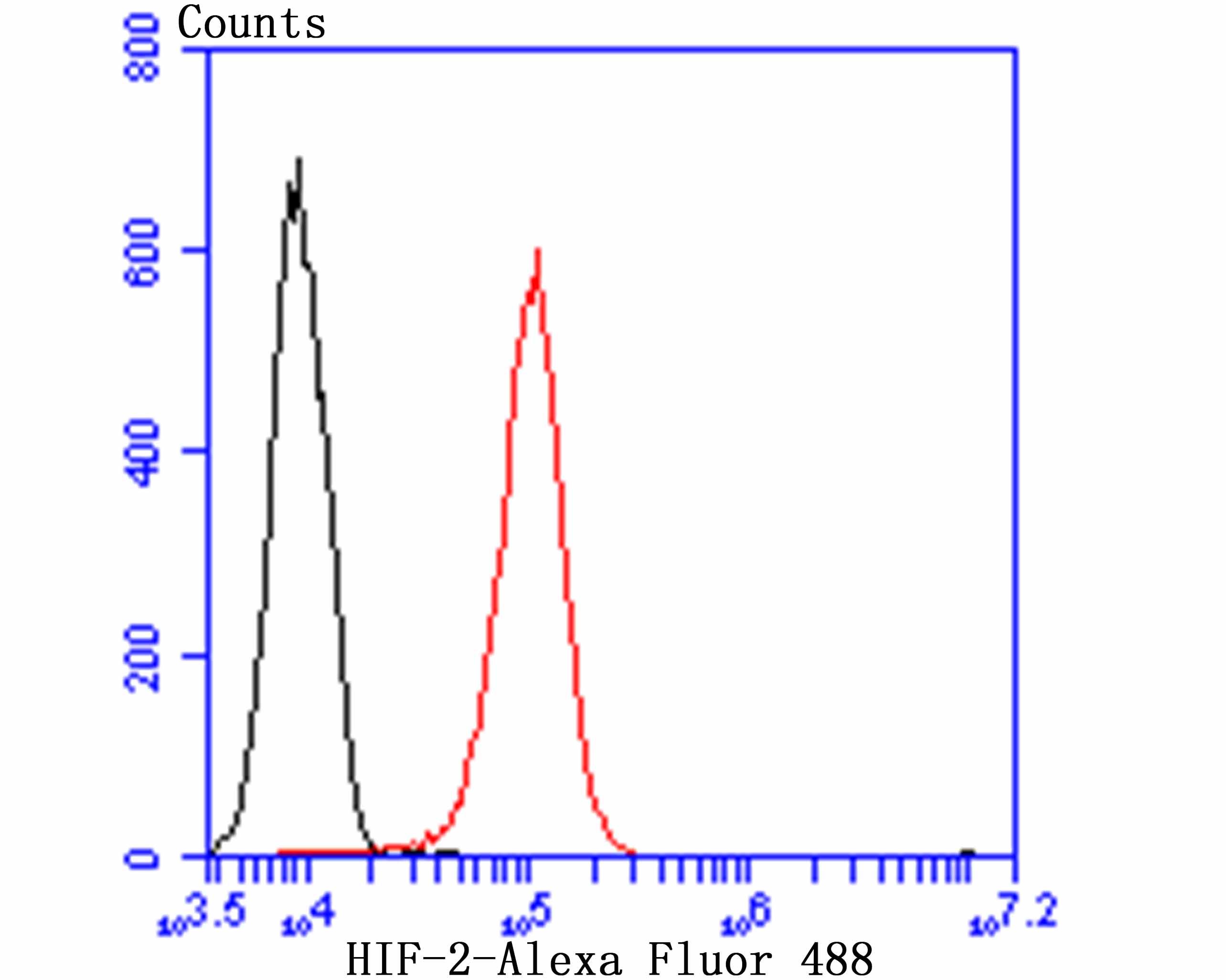
Fig6: Flow cytometric analysis of HUVEC cells with HIF-2 antibody at 1/100 dilution (red) compared with an unlabelled control (cells without incubation with primary antibody; black). Alexa Fluor 488-conjugated goat anti rabbit IgG was used as the secondary antibody.| Product Name | HIF-2 alpha [JB24-42] |
|---|---|
| Antibody Type | Primary Antibodies |
| Antigen Alias | Basic helix loop helix PAS protein MOP2 antibody Basic-helix-loop-helix-PAS protein MOP2 antibody bHLHe73 antibody Class E basic helix-loop-helix protein 73 antibody ECYT4 antibody Endothelial PAS domain containing protein 1 antibody Endothelial pas domain protein 1 antibody Endothelial PAS domain-containing protein 1 antibody EPAS 1 antibody EPAS-1 antibody EPAS1 antibody EPAS1_HUMAN antibody HIF 1 alpha like factor antibody HIF 2 alpha antibody HIF-1-alpha-like factor antibody HIF-2-alpha antibody HIF2-alpha antibody HIF2A antibody HLF antibody Hypoxia inducible factor 2 alpha antibody Hypoxia inducible factor 2 alpha subunit antibody Hypoxia-inducible factor 2-alpha antibody Member of PAS protein 2 antibody Member of pas superfamily 2 antibody MOP 2 antibody MOP2 antibody PAS domain-containing protein 2 antibody PASD2 antibody |
| Product description | Cell growth and viability is compromised by oxygen deprivation (hypoxia). Hypoxia-inducible factors, including HIF-1α, HIF-1β (also designated Arnt 1), EPAS-1 (also designated HIF-2α) and HIF-3α, induce glycolysis, erythropoiesis and angiogenesis in order to restore oxygen homeostasis. Hypoxia-inducible factors are members of the Per-Arnt-Sim (PAS) domain transcription factor family. In response to hypoxia, HIF-1α is upregulated and forms a heterodimer with Arnt 1 to form the HIF-1 complex. The HIF-1 complex recognizes and binds to the hypoxia responsive element (HRE) of hypoxia-inducible genes, thereby activating transcription. Hypoxia-inducible expression of some genes such as Glut-1, p53, p21 or Bcl-2, is HIF-1α dependent, whereas expression of others, such as p27, GADD 153 or HO-1, is HIF-1α independent. EPAS-1 and HIF-3α have also been shown to form heterodimeric complexes with Arnt 1 in response to hypoxia. |
| Immunogen | Recombinant protein |
| Clonality | Monoclonal |
|---|---|
| Isotype | IgG |
| Host Species | Recombinant rabbit |
| Tested Applications | WBIHCFC |
| WB:1:500-1:2,000 IHC:1:50-1:200 FC:1:50-1:100 |
|
| Species Reactivity | HumanMouseRat |
| Concentration | 1mg/ml |
| Alternative Names | Basic helix loop helix PAS protein MOP2 antibody Basic-helix-loop-helix-PAS protein MOP2 antibody bHLHe73 antibody Class E basic helix-loop-helix protein 73 antibody ECYT4 antibody Endothelial PAS domain containing protein 1 antibody Endothelial pas domain protein 1 antibody Endothelial PAS domain-containing protein 1 antibody EPAS 1 antibody EPAS-1 antibody EPAS1 antibody EPAS1_HUMAN antibody HIF 1 alpha like factor antibody HIF 2 alpha antibody HIF-1-alpha-like factor antibody HIF-2-alpha antibody HIF2-alpha antibody HIF2A antibody HLF antibody Hypoxia inducible factor 2 alpha antibody Hypoxia inducible factor 2 alpha subunit antibody Hypoxia-inducible factor 2-alpha antibody Member of PAS protein 2 antibody Member of pas superfamily 2 antibody MOP 2 antibody MOP2 antibody PAS domain-containing protein 2 antibody PASD2 antibody |
|---|---|
| Molecular Weight(MW) | 96 kDa |
| Cellular Localization | Nucleus. |
Application
Fig1: Western blot analysis of HIF-2 alpha on SiHa cell lysate using anti-HIF-2 alpha antibody at 1/500 dilution.
Application
Fig2: Immunohistochemical analysis of paraffin-embedded rat lung tissue using anti-HIF-2 alpha antibody. Counter stained with hematoxylin.
Application
Fig3: Immunohistochemical analysis of paraffin-embedded human breast cancer tissue using anti-HIF-2 alpha antibody. Counter stained with hematoxylin.
Application
Fig4: Immunohistochemical analysis of paraffin-embedded human placenta tissue using anti-HIF-2 alpha antibody. Counter stained with hematoxylin.
Application
Fig5: Immunohistochemical analysis of paraffin-embedded mouse colon tissue using anti-HIF-2 alpha antibody. Counter stained with hematoxylin.
Application
Fig6: Flow cytometric analysis of HUVEC cells with HIF-2 antibody at 1/100 dilution (red) compared with an unlabelled control (cells without incubation with primary antibody; black). Alexa Fluor 488-conjugated goat anti rabbit IgG was used as the secondary antibody.| Positive Control | SiHa, rat lung tissue, human breast cancer tissue, human placenta tissue, mouse colon tissue, HUVEC. |
|---|---|
| Application Notes | WB:1:500-1:2,000 IHC:1:50-1:200 FC:1:50-1:100 |
| Form | Liquid |
|---|---|
| Storage Instructions | Store at +4℃ after thawing. Aliquot store at -20℃ or -80℃. Avoid repeated freeze / thaw cycles. |
| Storage Buffer | 1*TBS (pH7.4), 1%BSA, 40%Glycerol. Preservative: 0.05% Sodium Azide. |


