Application
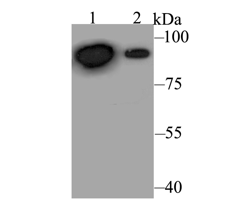
Fig1: Western blot analysis of Perforin on different lysates. Proteins were transferred to a PVDF membrane and blocked with 5% BSA in PBS for 1 hour at room temperature. The primary antibody was used at a 1:500 dilution in 5% BSA at room temperature for 2 hours. Goat Anti-Rabbit IgG - HRP Secondary Antibody (HA1001) at 1:5,000 dilution was used for 1 hour at room temperature. Positive control: Lane 1: HepG2 cell lysate Lane 2: Daudi cell lysateApplication
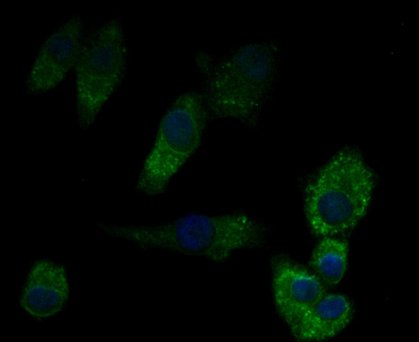
Fig2: ICC staining Perforin in A549 cells (green). Formalin fixed cells were permeabilized with 0.1% Triton X-100 in TBS for 10 minutes at room temperature and blocked with 1% Blocker BSA for 15 minutes at room temperature. Cells were probed with Perforin polyclonal antibody at a dilution of 1:200 for 1 hour at room temperature, washed with PBS. Alexa Fluorc™ 488 Goat anti-Rabbit IgG was used as the secondary antibody at 1/100 dilution. The nuclear counter stain is DAPI (blue).Application
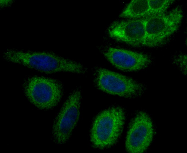
Fig3: ICC staining Perforin in HepG2 cells (green). Formalin fixed cells were permeabilized with 0.1% Triton X-100 in TBS for 10 minutes at room temperature and blocked with 1% Blocker BSA for 15 minutes at room temperature. Cells were probed with Perforin polyclonal antibody at a dilution of 1:200 for 1 hour at room temperature, washed with PBS. Alexa Fluorc™ 488 Goat anti-Rabbit IgG was used as the secondary antibody at 1/100 dilution. The nuclear counter stain is DAPI (blue).Application
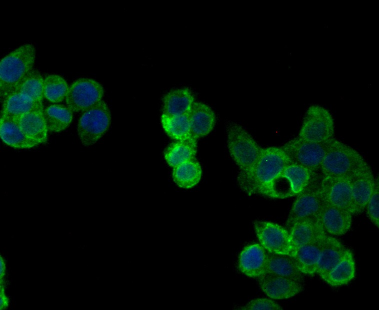
Fig4: ICC staining Perforin in HT-29 cells (green). Formalin fixed cells were permeabilized with 0.1% Triton X-100 in TBS for 10 minutes at room temperature and blocked with 1% Blocker BSA for 15 minutes at room temperature. Cells were probed with Perforin polyclonal antibody at a dilution of 1:200 for 1 hour at room temperature, washed with PBS. Alexa Fluorc™ 488 Goat anti-Rabbit IgG was used as the secondary antibody at 1/100 dilution. The nuclear counter stain is DAPI (blue).Application
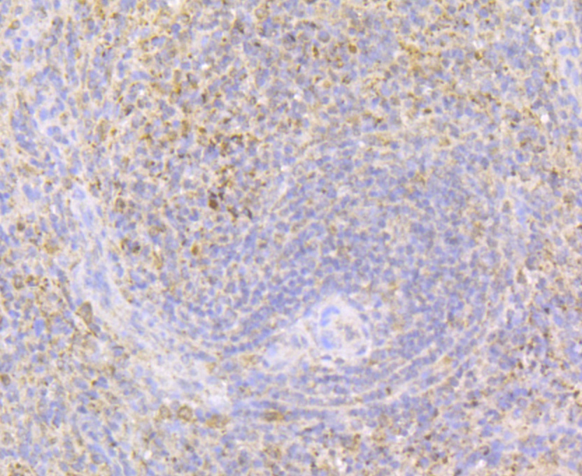
Fig5: Immunohistochemical analysis of paraffin-embedded human spleen tissue using anti-Perforin antibody. The section was pre-treated using heat mediated antigen retrieval with sodium citrate buffer (pH 6.0) for 20 minutes. The tissues were blocked in 5% BSA for 30 minutes at room temperature, washed with ddH2O and PBS, and then probed with the antibody (ER1803-77) at 1/200 dilution, for 30 minutes at room temperature and detected using an HRP conjugated compact polymer system. DAB was used as the chrogen. Counter stained with hematoxylin and mounted with DPX.Application
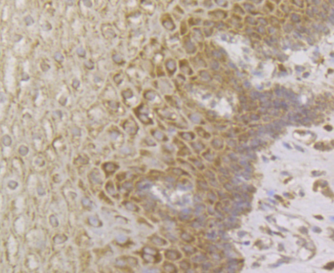
Fig6: Immunohistochemical analysis of paraffin-embedded human esophagus tissue using anti-Perforin antibody. The section was pre-treated using heat mediated antigen retrieval with sodium citrate buffer (pH 6.0) for 20 minutes. The tissues were blocked in 5% BSA for 30 minutes at room temperature, washed with ddH2O and PBS, and then probed with the antibody (ER1803-77) at 1/200 dilution, for 30 minutes at room temperature and detected using an HRP conjugated compact polymer system. DAB was used as the chrogen. Counter stained with hematoxylin and mounted with DPX.Application
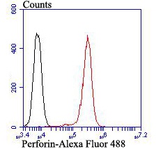
Fig7: Flow cytometric analysis of Perforin was done on HepG2 cells. The cells were fixed, permeabilized and stained with Perforin antibody at 1/100 dilution (red) compared with an unlabelled control (cells without incubation with primary antibody; black). After incubation of the primary antibody on room temperature for an hour, the cells was stained with a Alexa Fluor™ 488-conjugated goat anti-rabbit IgG Secondary antibody at 1/500 dilution for 30 minutes.| Product Name | Perforin |
|---|---|
| Antibody Type | Primary Antibodies |
| Antigen Alias | Cytolysin antibody FLH2 antibody HPLH2 antibody Lymphocyte pore-forming protein antibody P1 antibody PERF_HUMAN antibody perforin 1 (pore forming protein) antibody Perforin 1 antibody Perforin-1 antibody PFP antibody PGFL antibody PIGF antibody PIGF-2 antibody PLGF antibody Pore forming protein antibody prf1 antibody SHGC-10760 antibody |
| Product description | The protein encoded by this gene has structural and functional similarities to complement component 9 (C9). Like C9, this protein creates transmembrane tubules and is capable of lysing non-specifically a variety of target cells. Plays a key role in secretory granule-dependent cell death, and in defense against virus-infected or neoplastic cells. This protein is one of the main cytolytic proteins of cytolytic granules, and it is known to be a key effector molecule for T-cell- and natural killer-cell-mediated cytolysis. Defects in this gene cause familial hemophagocytic lymphohistiocytosis type 2 (HPLH2), a rare and lethal autosomal recessive disorder of early childhood. Alternative splicing results in multiple transcript variants encoding the same protein. Plays an important role in killing other cells that are recognized as non-self by the immune system, e.g. in transplant rejection or some forms of autoimmune disease. Can insert into the membrane of target cells in its calcium-bound form, oligomerize and form large pores. |
| Immunogen | Recombinant protein within Human Perforin aa 300-500. |
| Clonality | Polyclonal |
|---|---|
| Isotype | IgG |
| Host Species | Rabbit |
| Tested Applications | WBICCIHCFC |
| WB:1:500-1:2,000: ICC:1:50-1:200: IHC:1:50-1:200: FC:1:50-1:100: |
|
| Species Reactivity | Human |
| Concentration | 2mg/ml |
| Alternative Names | Cytolysin antibody FLH2 antibody HPLH2 antibody Lymphocyte pore-forming protein antibody P1 antibody PERF_HUMAN antibody perforin 1 (pore forming protein) antibody Perforin 1 antibody Perforin-1 antibody PFP antibody PGFL antibody PIGF antibody PIGF-2 antibody PLGF antibody Pore forming protein antibody prf1 antibody SHGC-10760 antibody |
|---|---|
| Molecular Weight(MW) | 90 kDa, predicted band size 61 kDa |
| Cellular Localization | Cell membrane, Endosome, Membrane, Secreted. |
| SwissProt ID | P14222 |
|---|
Application
Fig1: Western blot analysis of Perforin on different lysates. Proteins were transferred to a PVDF membrane and blocked with 5% BSA in PBS for 1 hour at room temperature. The primary antibody was used at a 1:500 dilution in 5% BSA at room temperature for 2 hours. Goat Anti-Rabbit IgG - HRP Secondary Antibody (HA1001) at 1:5,000 dilution was used for 1 hour at room temperature. Positive control: Lane 1: HepG2 cell lysate Lane 2: Daudi cell lysate
Application
Fig2: ICC staining Perforin in A549 cells (green). Formalin fixed cells were permeabilized with 0.1% Triton X-100 in TBS for 10 minutes at room temperature and blocked with 1% Blocker BSA for 15 minutes at room temperature. Cells were probed with Perforin polyclonal antibody at a dilution of 1:200 for 1 hour at room temperature, washed with PBS. Alexa Fluorc™ 488 Goat anti-Rabbit IgG was used as the secondary antibody at 1/100 dilution. The nuclear counter stain is DAPI (blue).
Application
Fig3: ICC staining Perforin in HepG2 cells (green). Formalin fixed cells were permeabilized with 0.1% Triton X-100 in TBS for 10 minutes at room temperature and blocked with 1% Blocker BSA for 15 minutes at room temperature. Cells were probed with Perforin polyclonal antibody at a dilution of 1:200 for 1 hour at room temperature, washed with PBS. Alexa Fluorc™ 488 Goat anti-Rabbit IgG was used as the secondary antibody at 1/100 dilution. The nuclear counter stain is DAPI (blue).
Application
Fig4: ICC staining Perforin in HT-29 cells (green). Formalin fixed cells were permeabilized with 0.1% Triton X-100 in TBS for 10 minutes at room temperature and blocked with 1% Blocker BSA for 15 minutes at room temperature. Cells were probed with Perforin polyclonal antibody at a dilution of 1:200 for 1 hour at room temperature, washed with PBS. Alexa Fluorc™ 488 Goat anti-Rabbit IgG was used as the secondary antibody at 1/100 dilution. The nuclear counter stain is DAPI (blue).
Application
Fig5: Immunohistochemical analysis of paraffin-embedded human spleen tissue using anti-Perforin antibody. The section was pre-treated using heat mediated antigen retrieval with sodium citrate buffer (pH 6.0) for 20 minutes. The tissues were blocked in 5% BSA for 30 minutes at room temperature, washed with ddH2O and PBS, and then probed with the antibody (ER1803-77) at 1/200 dilution, for 30 minutes at room temperature and detected using an HRP conjugated compact polymer system. DAB was used as the chrogen. Counter stained with hematoxylin and mounted with DPX.
Application
Fig6: Immunohistochemical analysis of paraffin-embedded human esophagus tissue using anti-Perforin antibody. The section was pre-treated using heat mediated antigen retrieval with sodium citrate buffer (pH 6.0) for 20 minutes. The tissues were blocked in 5% BSA for 30 minutes at room temperature, washed with ddH2O and PBS, and then probed with the antibody (ER1803-77) at 1/200 dilution, for 30 minutes at room temperature and detected using an HRP conjugated compact polymer system. DAB was used as the chrogen. Counter stained with hematoxylin and mounted with DPX.
Application
Fig7: Flow cytometric analysis of Perforin was done on HepG2 cells. The cells were fixed, permeabilized and stained with Perforin antibody at 1/100 dilution (red) compared with an unlabelled control (cells without incubation with primary antibody; black). After incubation of the primary antibody on room temperature for an hour, the cells was stained with a Alexa Fluor™ 488-conjugated goat anti-rabbit IgG Secondary antibody at 1/500 dilution for 30 minutes.| Positive Control | HepG2, Daudi, A549, HT-29, human spleen tissue, human esophagus tissue. |
|---|---|
| Application Notes | WB:1:500-1:2,000: ICC:1:50-1:200: IHC:1:50-1:200: FC:1:50-1:100: |
| Form | Liquid |
|---|---|
| Storage Instructions | Store at +4℃ after thawing. Aliquot store at -20℃. Avoid repeated freeze / thaw cycles. |
| Storage Buffer | 1*TBS (pH7.4), 1%BSA, 50%Glycerol. Preservative: 0.05% Sodium Azide. |


