Application
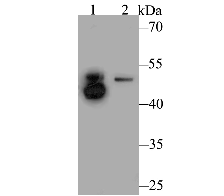
Fig1: Western blot analysis of Integrin linked ILK on different lysates. Proteins were transferred to a PVDF membrane and blocked with 5% BSA in PBS for 1 hour at room temperature. The primary antibody was used at a 1:500 dilution in 5% BSA at room temperature for 2 hours. Goat Anti-Rabbit IgG - HRP Secondary Antibody (HA1001) at 1:5,000 dilution was used for 1 hour at room temperature. Positive control: Lane 1: Mouse skeletal muscle tissue lysate Lane 2: Rat lung tissue lysateApplication
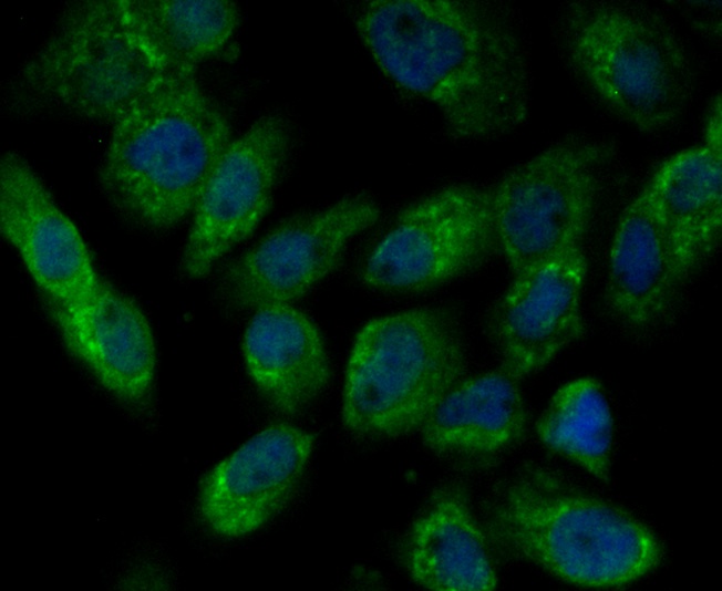
Fig2: ICC staining Integrin linked ILK in A431 cells (green). Formalin fixed cells were permeabilized with 0.1% Triton X-100 in TBS for 10 minutes at room temperature and blocked with 1% Blocker BSA for 15 minutes at room temperature. Cells were probed with Integrin linked ILK polyclonal antibody at a dilution of 1:100 for 1 hour at room temperature, washed with PBS. Alexa Fluorc™ 488 Goat anti-Rabbit IgG was used as the secondary antibody at 1/100 dilution. The nuclear counter stain is DAPI (blue).Application
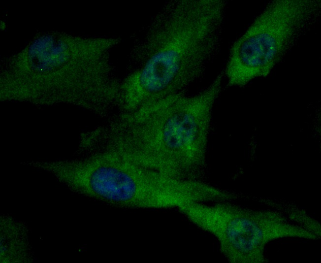
Fig3: ICC staining Integrin linked ILK in MG-63 cells (green). Formalin fixed cells were permeabilized with 0.1% Triton X-100 in TBS for 10 minutes at room temperature and blocked with 1% Blocker BSA for 15 minutes at room temperature. Cells were probed with Integrin linked ILK polyclonal antibody at a dilution of 1:50 for 1 hour at room temperature, washed with PBS. Alexa Fluorc™ 488 Goat anti-Rabbit IgG was used as the secondary antibody at 1/100 dilution. The nuclear counter stain is DAPI (blue).Application
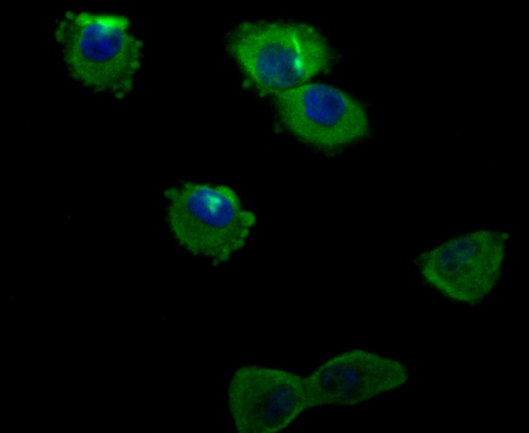
Fig4: ICC staining Integrin linked ILK in PANC-1 cells (green). Formalin fixed cells were permeabilized with 0.1% Triton X-100 in TBS for 10 minutes at room temperature and blocked with 1% Blocker BSA for 15 minutes at room temperature. Cells were probed with Integrin linked ILK polyclonal antibody at a dilution of 1:50 for 1 hour at room temperature, washed with PBS. Alexa Fluorc™ 488 Goat anti-Rabbit IgG was used as the secondary antibody at 1/100 dilution. The nuclear counter stain is DAPI (blue).Application
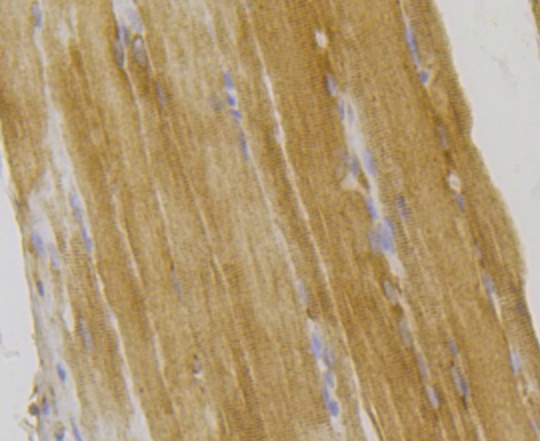
Fig5: Immunohistochemical analysis of paraffin-embedded rat skeletal muscle tissue using anti-Integrin linked ILK antibody. The section was pre-treated using heat mediated antigen retrieval with Tris-EDTA buffer (pH 8.0-8.4) for 20 minutes.The tissues were blocked in 5% BSA for 30 minutes at room temperature, washed with ddH2O and PBS, and then probed with the antibody (ER1803-78) at 1/200 dilution, for 30 minutes at room temperature and detected using an HRP conjugated compact polymer system. DAB was used as the chrogen. Counter stained with hematoxylin and mounted with DPX.Application
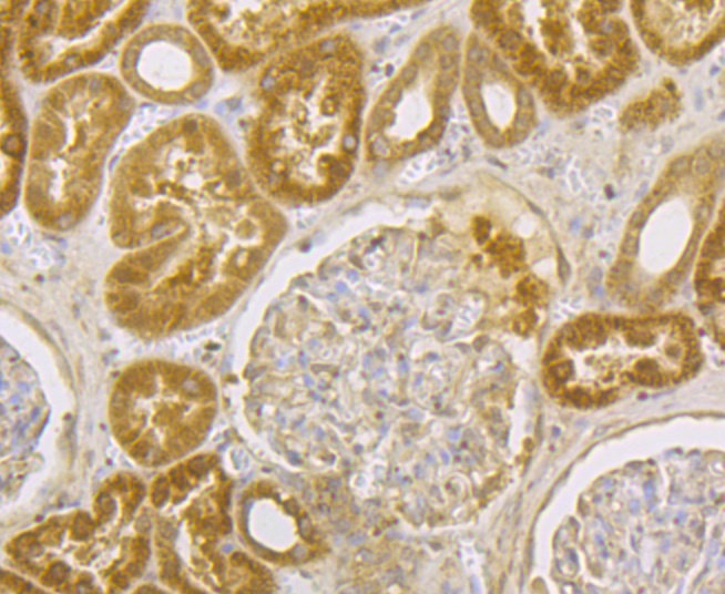
Fig6: Immunohistochemical analysis of paraffin-embedded human kidney tissue using anti-Integrin linked ILK antibody. The section was pre-treated using heat mediated antigen retrieval with Tris-EDTA buffer (pH 8.0-8.4) for 20 minutes.The tissues were blocked in 5% BSA for 30 minutes at room temperature, washed with ddH2O and PBS, and then probed with the antibody (ER1803-78) at 1/200 dilution, for 30 minutes at room temperature and detected using an HRP conjugated compact polymer system. DAB was used as the chrogen. Counter stained with hematoxylin and mounted with DPX.Application
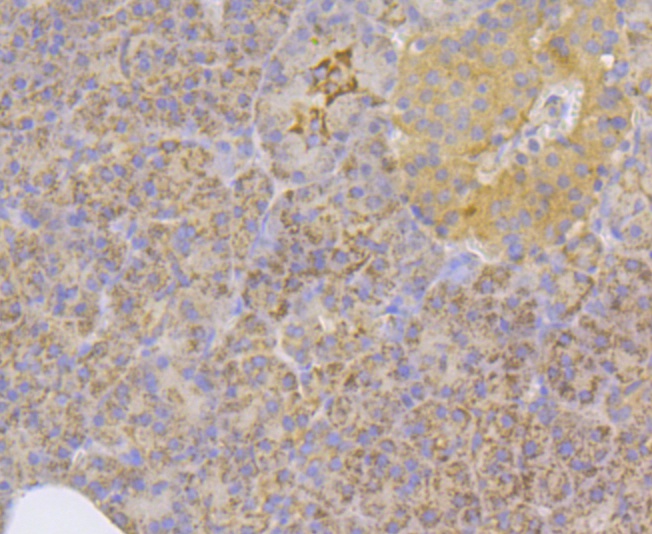
Fig7: Immunohistochemical analysis of paraffin-embedded human pancreas tissue using anti-Integrin linked ILK antibody. The section was pre-treated using heat mediated antigen retrieval with Tris-EDTA buffer (pH 8.0-8.4) for 20 minutes.The tissues were blocked in 5% BSA for 30 minutes at room temperature, washed with ddH2O and PBS, and then probed with the antibody (ER1803-78) at 1/200 dilution, for 30 minutes at room temperature and detected using an HRP conjugated compact polymer system. DAB was used as the chrogen. Counter stained with hematoxylin and mounted with DPX.Application
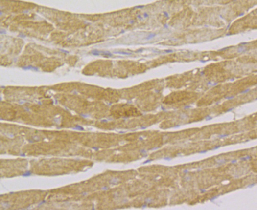
Fig8: Immunohistochemical analysis of paraffin-embedded mouse heart tissue using anti-Integrin linked ILK antibody. The section was pre-treated using heat mediated antigen retrieval with Tris-EDTA buffer (pH 8.0-8.4) for 20 minutes.The tissues were blocked in 5% BSA for 30 minutes at room temperature, washed with ddH2O and PBS, and then probed with the antibody (ER1803-78) at 1/50 dilution, for 30 minutes at room temperature and detected using an HRP conjugated compact polymer system. DAB was used as the chrogen. Counter stained with hematoxylin and mounted with DPX.| Product Name | Integrin linked ILK |
|---|---|
| Antibody Type | Primary Antibodies |
| Antigen Alias | 59 kDa serine/threonine protein kinase antibody 59 kDa serine/threonine-protein kinase antibody DKFZp686F1765 antibody Epididymis secretory protein Li 28 antibody HEL S 28 antibody ILK 1 antibody ILK 2 antibody ILK antibody ILK-1 antibody ILK-2 antibody ILK_HUMAN antibody ILK1 antibody ILK2 antibody Integrin linked kinase 2 antibody Integrin linked Kinase antibody Integrin linked protein kinase antibody Integrin-linked protein kinase antibody p59 antibody p59ILK antibody |
| Product description | Integrins are heterodimers composed of non-covalently associated transmembrane a and b subunits. The 16 a and 8 b subunits heterodimerize to produce more than 20 different receptors. Most integrin receptors bind to ligands that are components of the extracellular matrix. Certain integrins can also bind to soluble ligands such as Fibrinogen, or to counterreceptors on adjacent cells, such as the intracellular adhesion molecules (ICAMs), leading to aggregation of cells. In addition to mediating cell adhesion and cytoskeletal organization, integrins function as signaling receptors. Signals transduced by integrins play a role in many biological processes, including cell growth, differentiation, migration and apoptosis. ILK (integrin-linked kinase) was identified as a serine/threonine kinase that phosphorylates b1 and b3 integrins. ILK expression has been shown to be reduced in response to Fibronectin, a known integrin ligand. Overexpression of ILK was shown to upregulate the Fibronectin matrix assembly in epithelial cells, indicating a potential role for ILK in cell growth, cell survival and tumorigenesis. |
| Immunogen | Recombinant protein within Human Integrin linked ILK aa 1-290. |
| Clonality | Polyclonal |
|---|---|
| Isotype | IgG |
| Host Species | Rabbit |
| Tested Applications | WBICCIHC |
| WB:1:500: ICC:1:50-1:200: IHC:1:50-1:200: |
|
| Species Reactivity | HumanMouseRat |
| Concentration | 1mg/ml |
| Alternative Names | 59 kDa serine/threonine protein kinase antibody 59 kDa serine/threonine-protein kinase antibody DKFZp686F1765 antibody Epididymis secretory protein Li 28 antibody HEL S 28 antibody ILK 1 antibody ILK 2 antibody ILK antibody ILK-1 antibody ILK-2 antibody ILK_HUMAN antibody ILK1 antibody ILK2 antibody Integrin linked kinase 2 antibody Integrin linked Kinase antibody Integrin linked protein kinase antibody Integrin-linked protein kinase antibody p59 antibody p59ILK antibody |
|---|---|
| Molecular Weight(MW) | 51 kDa |
| Cellular Localization | Cell junction, Cell membrane, Cytoplasm. |
Application
Fig1: Western blot analysis of Integrin linked ILK on different lysates. Proteins were transferred to a PVDF membrane and blocked with 5% BSA in PBS for 1 hour at room temperature. The primary antibody was used at a 1:500 dilution in 5% BSA at room temperature for 2 hours. Goat Anti-Rabbit IgG - HRP Secondary Antibody (HA1001) at 1:5,000 dilution was used for 1 hour at room temperature. Positive control: Lane 1: Mouse skeletal muscle tissue lysate Lane 2: Rat lung tissue lysate
Application
Fig2: ICC staining Integrin linked ILK in A431 cells (green). Formalin fixed cells were permeabilized with 0.1% Triton X-100 in TBS for 10 minutes at room temperature and blocked with 1% Blocker BSA for 15 minutes at room temperature. Cells were probed with Integrin linked ILK polyclonal antibody at a dilution of 1:100 for 1 hour at room temperature, washed with PBS. Alexa Fluorc™ 488 Goat anti-Rabbit IgG was used as the secondary antibody at 1/100 dilution. The nuclear counter stain is DAPI (blue).
Application
Fig3: ICC staining Integrin linked ILK in MG-63 cells (green). Formalin fixed cells were permeabilized with 0.1% Triton X-100 in TBS for 10 minutes at room temperature and blocked with 1% Blocker BSA for 15 minutes at room temperature. Cells were probed with Integrin linked ILK polyclonal antibody at a dilution of 1:50 for 1 hour at room temperature, washed with PBS. Alexa Fluorc™ 488 Goat anti-Rabbit IgG was used as the secondary antibody at 1/100 dilution. The nuclear counter stain is DAPI (blue).
Application
Fig4: ICC staining Integrin linked ILK in PANC-1 cells (green). Formalin fixed cells were permeabilized with 0.1% Triton X-100 in TBS for 10 minutes at room temperature and blocked with 1% Blocker BSA for 15 minutes at room temperature. Cells were probed with Integrin linked ILK polyclonal antibody at a dilution of 1:50 for 1 hour at room temperature, washed with PBS. Alexa Fluorc™ 488 Goat anti-Rabbit IgG was used as the secondary antibody at 1/100 dilution. The nuclear counter stain is DAPI (blue).
Application
Fig5: Immunohistochemical analysis of paraffin-embedded rat skeletal muscle tissue using anti-Integrin linked ILK antibody. The section was pre-treated using heat mediated antigen retrieval with Tris-EDTA buffer (pH 8.0-8.4) for 20 minutes.The tissues were blocked in 5% BSA for 30 minutes at room temperature, washed with ddH2O and PBS, and then probed with the antibody (ER1803-78) at 1/200 dilution, for 30 minutes at room temperature and detected using an HRP conjugated compact polymer system. DAB was used as the chrogen. Counter stained with hematoxylin and mounted with DPX.
Application
Fig6: Immunohistochemical analysis of paraffin-embedded human kidney tissue using anti-Integrin linked ILK antibody. The section was pre-treated using heat mediated antigen retrieval with Tris-EDTA buffer (pH 8.0-8.4) for 20 minutes.The tissues were blocked in 5% BSA for 30 minutes at room temperature, washed with ddH2O and PBS, and then probed with the antibody (ER1803-78) at 1/200 dilution, for 30 minutes at room temperature and detected using an HRP conjugated compact polymer system. DAB was used as the chrogen. Counter stained with hematoxylin and mounted with DPX.
Application
Fig7: Immunohistochemical analysis of paraffin-embedded human pancreas tissue using anti-Integrin linked ILK antibody. The section was pre-treated using heat mediated antigen retrieval with Tris-EDTA buffer (pH 8.0-8.4) for 20 minutes.The tissues were blocked in 5% BSA for 30 minutes at room temperature, washed with ddH2O and PBS, and then probed with the antibody (ER1803-78) at 1/200 dilution, for 30 minutes at room temperature and detected using an HRP conjugated compact polymer system. DAB was used as the chrogen. Counter stained with hematoxylin and mounted with DPX.
Application
Fig8: Immunohistochemical analysis of paraffin-embedded mouse heart tissue using anti-Integrin linked ILK antibody. The section was pre-treated using heat mediated antigen retrieval with Tris-EDTA buffer (pH 8.0-8.4) for 20 minutes.The tissues were blocked in 5% BSA for 30 minutes at room temperature, washed with ddH2O and PBS, and then probed with the antibody (ER1803-78) at 1/50 dilution, for 30 minutes at room temperature and detected using an HRP conjugated compact polymer system. DAB was used as the chrogen. Counter stained with hematoxylin and mounted with DPX.| Positive Control | Mouse skeletal muscle tissue, rat lung tissue, A431, MG-63, PANC-1, rat skeletal muscle tissue, human kidney tissue, human pancreas tissue, mouse heart tissue. |
|---|---|
| Application Notes | WB:1:500: ICC:1:50-1:200: IHC:1:50-1:200: |
| Form | Liquid |
|---|---|
| Storage Instructions | Store at +4℃ after thawing. Aliquot store at -20℃. Avoid repeated freeze / thaw cycles. |
| Storage Buffer | 1*TBS (pH7.4), 1%BSA, 50%Glycerol. Preservative: 0.05% Sodium Azide. |


