Application
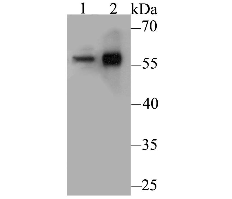
Fig1: Western blot analysis of CD4 on different lysates. Proteins were transferred to a PVDF membrane and blocked with 5% BSA in PBS for 1 hour at room temperature. The primary antibody was used at a 1:500 dilution in 5% BSA at room temperature for 2 hours. Goat Anti-Rabbit IgG - HRP Secondary Antibody (HA1001) at 1:5,000 dilution was used for 1 hour at room temperature. Positive control: Lane 1: U937 cell lysate Lane 2: Human thymus tissue lysateApplication
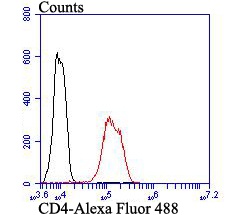
Fig2: Flow cytometric analysis of CD4 was done on Hela cells. The cells were fixed, permeabilized and stained with CD4 antibody at 1/100 dilution (red) compared with an unlabelled control (cells without incubation with primary antibody; black). After incubation of the primary antibody on room temperature for an hour, the cells was stained with a Alexa Fluor™ 488-conjugated goat anti-rabbit IgG Secondary antibody at 1/500 dilution for 30 minutes.| Product Name | CD4 |
|---|---|
| Antibody Type | Primary Antibodies |
| Antigen Alias | CD 4 antibody CD4 (L3T4) antibody CD4 antibody CD4 antigen (p55) antibody CD4 antigen antibody CD4 molecule antibody CD4 receptor antibody CD4+ Lymphocyte deficiency, included antibody CD4_HUMAN antibody CD4mut antibody L3T4 antibody Leu3 antibody Ly-4 antibody Lymphocyte antigen CD4 antibody MGC165891 antibody OTTHUMP00000238897 antibody p55 antibody T cell antigen T4 antibody T cell antigen T4/LEU3 antibody T cell differentiation antigen L3T4 antibody T cell OKT4 deficiency, included antibody T cell surface antigen T4/Leu 3 antibody T cell surface antigen T4/Leu3 antibody T cell surface glycoprotein CD4 antibody T-cell surface antigen T4/Leu-3 antibody T-cell surface glycoprotein CD4 antibody W3/25 antibody W3/25 antigen antibody |
| Product description | Integral membrane glycoprotein that plays an essential role in the immune response and serves multiple functions in responses against both external and internal offenses. In T-cells, functions primarily as a coreceptor for MHC class II molecule:peptide complex. The antigens presented by class II peptides are derived from extracellular proteins while class I peptides are derived from cytosolic proteins. Interacts simultaneously with the T-cell receptor (TCR) and the MHC class II presented by antigen presenting cells (APCs). In turn, recruits the Src kinase LCK to the vicinity of the TCR-CD3 complex. LCK then initiates different intracellular signaling pathways by phosphorylating various substrates ultimately leading to lymphokine production, motility, adhesion and activation of T-helper cells. In other cells such as macrophages or NK cells, plays a role in differentiation/activation, cytokine expression and cell migration in a TCR/LCK-independent pathway. Participates in the development of T-helper cells in the thymus and triggers the differentiation of monocytes into functional mature macrophages. |
| Immunogen | Recombinant protein with Human CD4 1-300 aa. |
| Clonality | Polyclonal |
|---|---|
| Isotype | IgG |
| Host Species | Rabbit |
| Tested Applications | WBFC |
| WB:1:500: FC:1:50-1:100: |
|
| Species Reactivity | Human |
| Concentration | 1mg/ml |
| Alternative Names | CD 4 antibody
CD4 (L3T4) antibody
CD4 antibody
CD4 antigen (p55) antibody
CD4 antigen antibody
CD4 molecule antibody
CD4 receptor antibody
CD4+ Lymphocyte deficiency included antibody CD4_HUMAN antibody CD4mut antibody L3T4 antibody Leu3 antibody Ly-4 antibody Lymphocyte antigen CD4 antibody MGC165891 antibody OTTHUMP00000238897 antibody p55 antibody T cell antigen T4 antibody T cell antigen T4/LEU3 antibody T cell differentiation antigen L3T4 antibody T cell OKT4 deficiency included antibody T cell surface antigen T4/Leu 3 antibody T cell surface antigen T4/Leu3 antibody T cell surface glycoprotein CD4 antibody T-cell surface antigen T4/Leu-3 antibody T-cell surface glycoprotein CD4 antibody W3/25 antibody W3/25 antigen antibody |
|---|---|
| Molecular Weight(MW) | 51 kDa |
| Cellular Localization | Cell Membrane. |
| SwissProt ID | P01730 |
|---|
Application
Fig1: Western blot analysis of CD4 on different lysates. Proteins were transferred to a PVDF membrane and blocked with 5% BSA in PBS for 1 hour at room temperature. The primary antibody was used at a 1:500 dilution in 5% BSA at room temperature for 2 hours. Goat Anti-Rabbit IgG - HRP Secondary Antibody (HA1001) at 1:5,000 dilution was used for 1 hour at room temperature. Positive control: Lane 1: U937 cell lysate Lane 2: Human thymus tissue lysate
Application
Fig2: Flow cytometric analysis of CD4 was done on Hela cells. The cells were fixed, permeabilized and stained with CD4 antibody at 1/100 dilution (red) compared with an unlabelled control (cells without incubation with primary antibody; black). After incubation of the primary antibody on room temperature for an hour, the cells was stained with a Alexa Fluor™ 488-conjugated goat anti-rabbit IgG Secondary antibody at 1/500 dilution for 30 minutes.| Positive Control | U937, human thymus tissue, Hela. |
|---|---|
| Application Notes | WB:1:500: FC:1:50-1:100: |
| Form | Liquid |
|---|---|
| Storage Instructions | Store at +4℃ after thawing. Aliquot store at -20℃. Avoid repeated freeze / thaw cycles. |
| Storage Buffer | 1*TBS (pH7.4), 1%BSA, 50%Glycerol. Preservative: 0.05% Sodium Azide. |


