Application
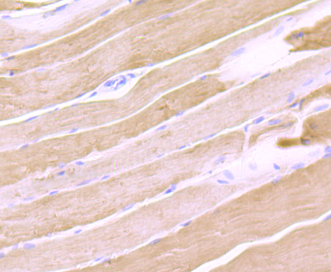
Fig1: Immunohistochemical analysis of paraffin-embedded rat skeletal muscle tissue using anti-Calsequestrin 1 antibody. Counter stained with hematoxylin.Application
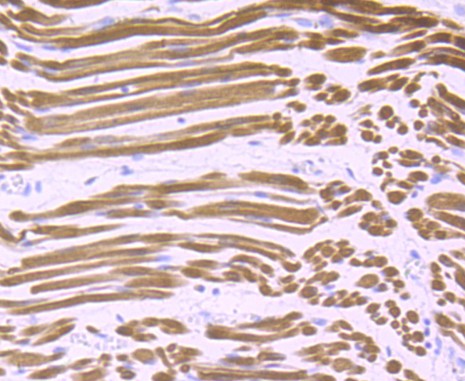
Fig2: Immunohistochemical analysis of paraffin-embedded human fetal skeletal muscle tissue using anti-Calsequestrin 1 antibody. Counter stained with hematoxylin.Application
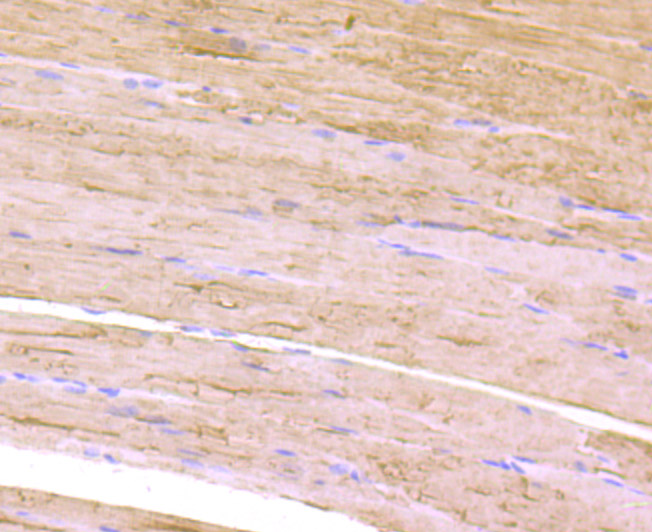
Fig3: Immunohistochemical analysis of paraffin-embedded mouse skeletal muscle tissue using anti-Calsequestrin 1 antibody. Counter stained with hematoxylin.Application
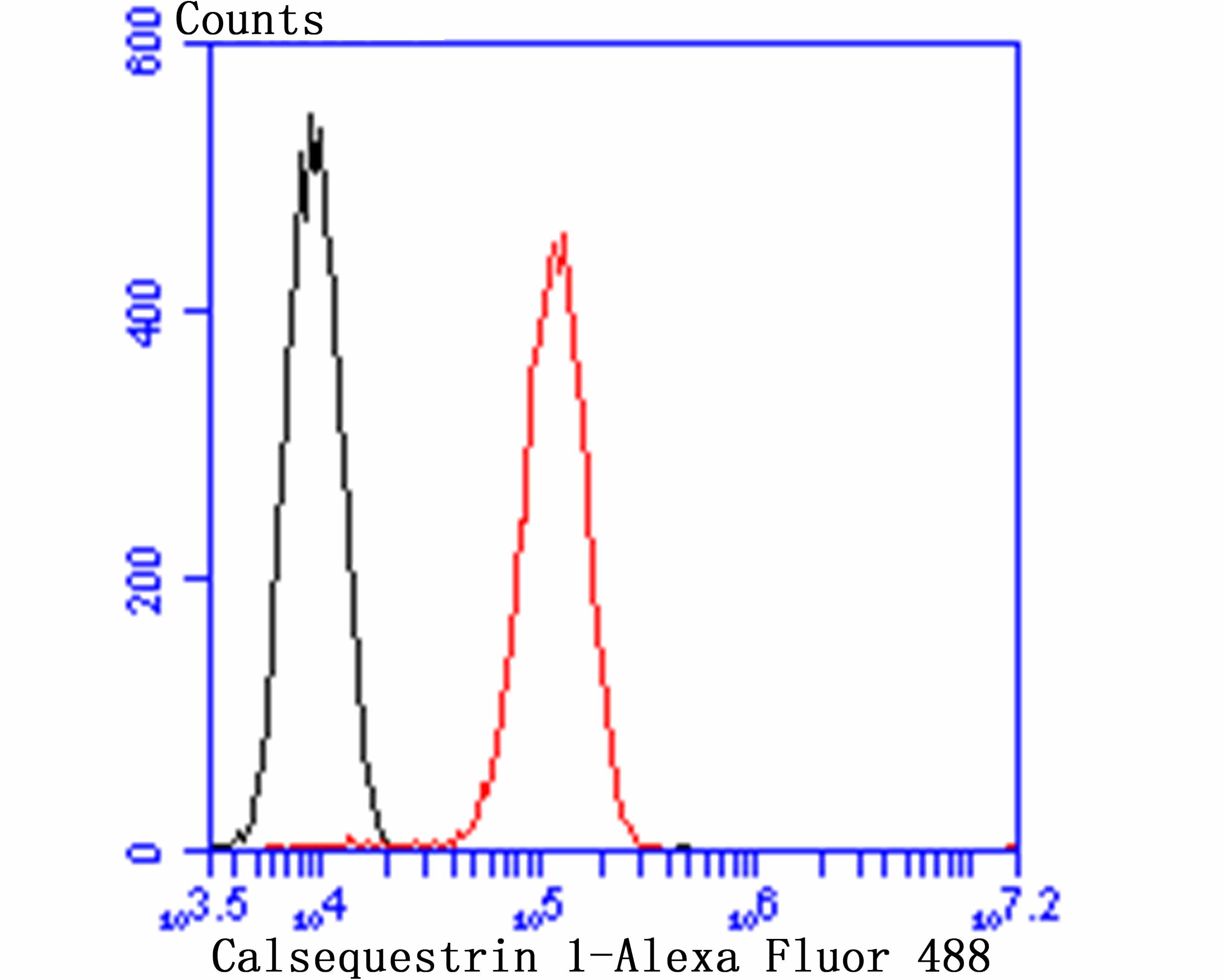
Fig4: Flow cytometric analysis of C2C12 cells with Calsequestrin 1 antibody at 1/100 dilution (red) compared with an unlabelled control (cells without incubation with primary antibody; black). Alexa Fluor 488-conjugated goat anti rabbit IgG was used as the secondary antibody.| Product Name | Calsequestrin 1 [JB57-37] |
|---|---|
| Antibody Type | Primary Antibodies |
| Antigen Alias | Calmitin antibody Calmitine antibody Calsequestrin 1 (fast twitch, skeletal muscle) antibody Calsequestrin antibody Calsequestrin, cell antibody Calsequestrin, skeletal muscle isoform antibody Calsequestrin-1 antibody CASQ antibody Casq1 antibody CASQ1_HUMAN antibody PDIB1 antibody skeletal muscle isoform antibody |
| Product description | Calsequestrin (CS, also known as CSQ) is the major calcium-binding protein of cardiac and skeletal muscle whose function is to sequester calcium in the lumen of the sarcoplasmic reticulum (SR). In mammals, there are two forms of this protein, calsequestrin 1 and calsequestrin 2, which encode fast-twitch skeletal muscle and cardiac calsequestrin, respectively. Calsequestrin 1, also known as Calmitin, is located in the terminal cisternae luminal spaces of the SR of fast skeletal muscle cells. Calsequestrin 2 is present in terminal cisternae luminal spaces of the SR of both cardiac and slow skeletal muscle cells. In addition, calsequestrin regulates the ryanodine receptor signaling (RyR) through Triadin and Junctin. |
| Immunogen | Recombinant protein |
| Clonality | Monoclonal |
|---|---|
| Isotype | IgG |
| Host Species | Recombinant rabbit |
| Tested Applications | WBIHCFC |
| WB:1:500 IHC:1:50-1:200 FC:1:50-1:100 |
|
| Species Reactivity | HumanMouseRat |
| Concentration | 1mg/ml |
| Alternative Names | Calmitin antibody
Calmitine antibody
Calsequestrin 1 (fast twitch skeletal muscle) antibody Calsequestrin antibody Calsequestrin cell antibody Calsequestrin skeletal muscle isoform antibody Calsequestrin-1 antibody CASQ antibody Casq1 antibody CASQ1_HUMAN antibody PDIB1 antibody skeletal muscle isoform antibody |
|---|---|
| Molecular Weight(MW) | 45 kDa |
| Cellular Localization | Endoplasmic reticulum. Mitochondrion. |
Application
Fig1: Immunohistochemical analysis of paraffin-embedded rat skeletal muscle tissue using anti-Calsequestrin 1 antibody. Counter stained with hematoxylin.
Application
Fig2: Immunohistochemical analysis of paraffin-embedded human fetal skeletal muscle tissue using anti-Calsequestrin 1 antibody. Counter stained with hematoxylin.
Application
Fig3: Immunohistochemical analysis of paraffin-embedded mouse skeletal muscle tissue using anti-Calsequestrin 1 antibody. Counter stained with hematoxylin.
Application
Fig4: Flow cytometric analysis of C2C12 cells with Calsequestrin 1 antibody at 1/100 dilution (red) compared with an unlabelled control (cells without incubation with primary antibody; black). Alexa Fluor 488-conjugated goat anti rabbit IgG was used as the secondary antibody.| Positive Control | Rat skeletal muscle tissue, mouse skeletal muscle tissue, human fetal skeletal muscle tissue, C2C12. |
|---|---|
| Application Notes | WB:1:500 IHC:1:50-1:200 FC:1:50-1:100 |
| Form | Liquid |
|---|---|
| Storage Instructions | Store at +4℃ after thawing. Aliquot store at -20℃ or -80℃. Avoid repeated freeze / thaw cycles. |
| Storage Buffer | 1*TBS (pH7.4), 1%BSA, 40%Glycerol. Preservative: 0.05% Sodium Azide. |


