Application
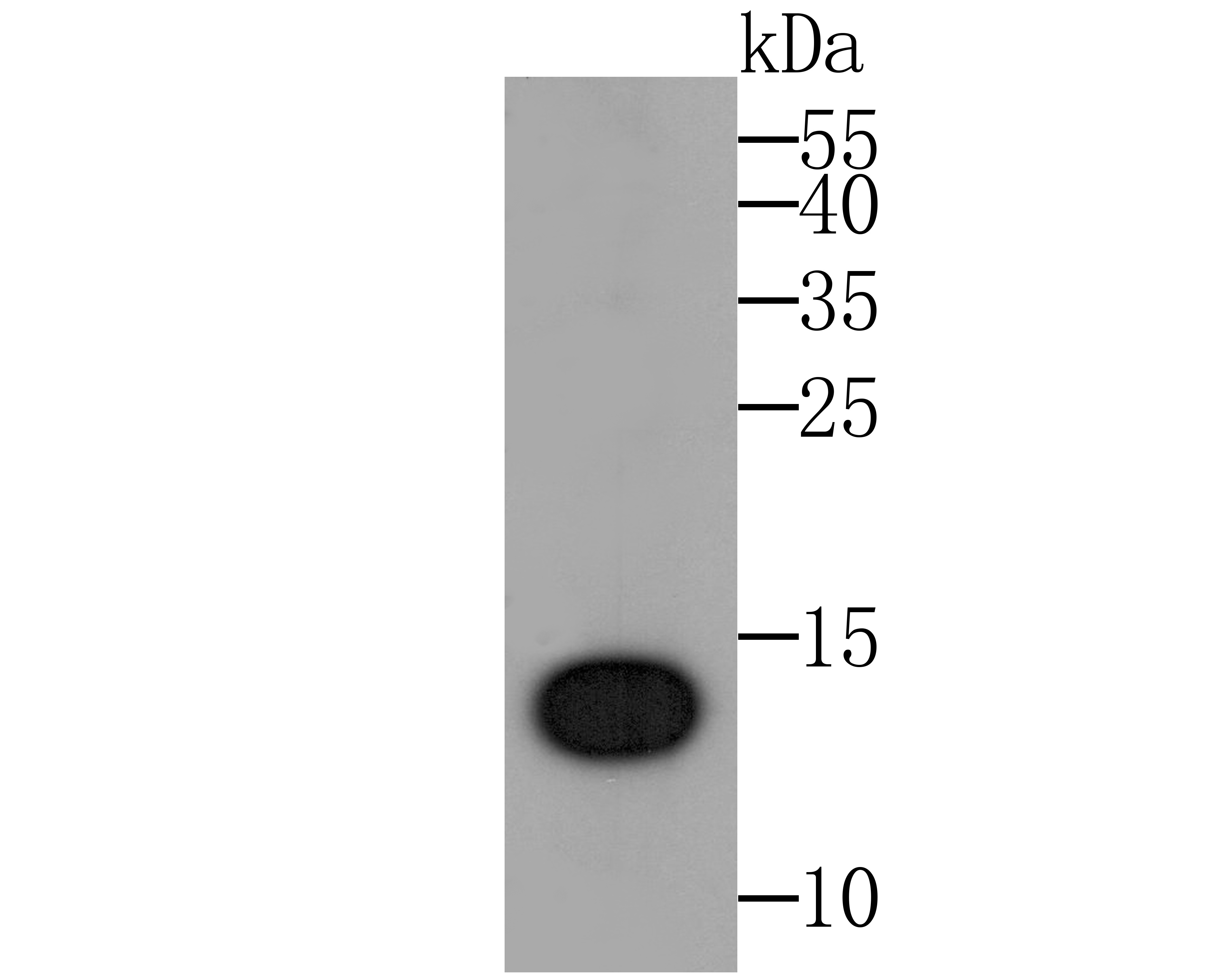
Fig1: Western blot analysis of IL-22 on IL22 recombinant protein. Proteins were transferred to a PVDF membrane and blocked with 5% BSA in PBS for 1 hour at room temperature. The primary antibody was used at a 1:500 dilution in 5% BSA at room temperature for 2 hours. Goat Anti-Rabbit IgG - HRP Secondary Antibody (HA1001) at 1:5,000 dilution was used for 1 hour at room temperature.Application
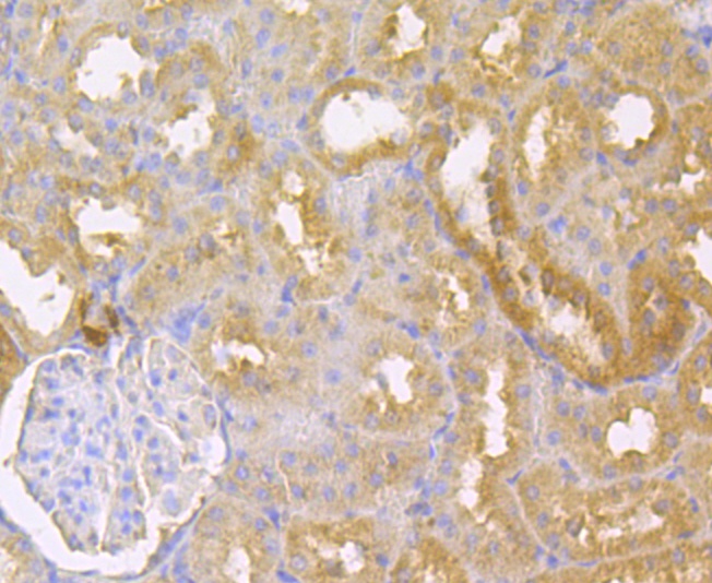
Fig2: Immunohistochemical analysis of paraffin-embedded rat kidney tissue using anti-IL-22 antibody. The section was pre-treated using heat mediated antigen retrieval with sodium citrate buffer (pH 6.0) for 20 minutes. The tissues were blocked in 5% BSA for 30 minutes at room temperature, washed with ddH2O and PBS, and then probed with the antibody (ER1803-85) at 1/200 dilution, for 30 minutes at room temperature and detected using an HRP conjugated compact polymer system. DAB was used as the chrogen. Counter stained with hematoxylin and mounted with DPX.Application
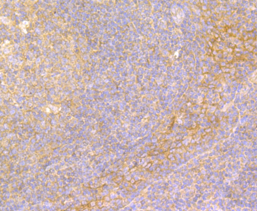
Fig3: Immunohistochemical analysis of paraffin-embedded human tonsil tissue using anti-IL-22 antibody. The section was pre-treated using heat mediated antigen retrieval with sodium citrate buffer (pH 6.0) for 20 minutes. The tissues were blocked in 5% BSA for 30 minutes at room temperature, washed with ddH2O and PBS, and then probed with the antibody (ER1803-85) at 1/200 dilution, for 30 minutes at room temperature and detected using an HRP conjugated compact polymer system. DAB was used as the chrogen. Counter stained with hematoxylin and mounted with DPX.Application
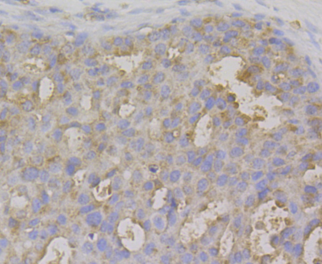
Fig4: Immunohistochemical analysis of paraffin-embedded human breasr cancer tissue using anti-IL-22 antibody. The section was pre-treated using heat mediated antigen retrieval with sodium citrate buffer (pH 6.0) for 20 minutes. The tissues were blocked in 5% BSA for 30 minutes at room temperature, washed with ddH2O and PBS, and then probed with the antibody (ER1803-85) at 1/200 dilution, for 30 minutes at room temperature and detected using an HRP conjugated compact polymer system. DAB was used as the chrogen. Counter stained with hematoxylin and mounted with DPX.Application
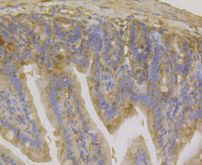
Fig5: Immunohistochemical analysis of paraffin-embedded mouse colon tissue using anti-IL-22 antibody. The section was pre-treated using heat mediated antigen retrieval with sodium citrate buffer (pH 6.0) for 20 minutes. The tissues were blocked in 5% BSA for 30 minutes at room temperature, washed with ddH2O and PBS, and then probed with the antibody (ER1803-85) at 1/200 dilution, for 30 minutes at room temperature and detected using an HRP conjugated compact polymer system. DAB was used as the chrogen. Counter stained with hematoxylin and mounted with DPX.| Product Name | IL-22 |
|---|---|
| Antibody Type | Primary Antibodies |
| Antigen Alias | Cytokine Zcyto18 antibody IL 10 related T cell derived inducible factor antibody IL 21 antibody IL 22 antibody IL D110 antibody IL TIF antibody IL-10-related T-cell-derived-inducible factor antibody IL-22 antibody IL-TIF antibody IL21 antibody Il22 antibody IL22_HUMAN antibody ILD110 antibody ILTIF antibody Interleukin 10 related T cell derived inducible factor antibody interleukin 21 antibody Interleukin 22 antibody Interleukin-22 antibody MGC79382 antibody MGC79384 antibody TIFa antibody TIFIL 23 antibody TIFIL23 antibody UNQ3099/PRO10096 antibody zcyto18 antibody |
| Product description | IL-22 a member of a group of cytokines called the IL-10 family or IL-10 superfamily (including IL-19, IL-20, IL-24, and IL-26), a class of potent mediators of cellular inflammatory responses. It shares use of IL-10R2 in cell signaling with other members of this family, IL-10, IL-26, IL-28A/B and IL-29. IL-22 is produced by activated NK and T cells and initiates innate immune responses against bacterial pathogens especially in epithelial cells such as respiratory and gut epithelial cells. IL-22 along with IL-17 is rapidly produced by splenic LTi-like cells and also produced by Th17 cells and likely plays a role in the coordinated response of both adaptive innate immune systems, autoimmunity and tissue regeneration. IL-22 biological activity is initiated by binding to a cell-surface complex composed of IL-22R1 and IL-10R2 receptor chains. IL-22 and IL-10 receptor chains play a role in cellular targeting and signal transduction to selectively initiate and regulate immune responses. IL-22 can contribute to immune disease through the stimulation of inflammatory responses, S100s and defensins. IL-22 also promotes hepatocyte survival in the liver and epithelial cells in the lung and gut similar to IL-10. In some contexts, the pro-inflammatory versus tissue-protective functions of IL-22 are regulated by the often co-expressed cytokine IL-17A. |
| Immunogen | Recombinant protein within human IL-22 aa 34-179. |
| Clonality | Polyclonal |
|---|---|
| Isotype | IgG |
| Host Species | Rabbit |
| Tested Applications | ICC/IFWBIHC |
| ELISA:1:5000-1:10,000: WB:1:500: IHC:1:50-1:200: |
|
| Species Reactivity | HumanMouseRat |
| Concentration | 1mg/ml |
| Alternative Names | Cytokine Zcyto18 antibody IL 10 related T cell derived inducible factor antibody IL 21 antibody IL 22 antibody IL D110 antibody IL TIF antibody IL-10-related T-cell-derived-inducible factor antibody IL-22 antibody IL-TIF antibody IL21 antibody Il22 antibody IL22_HUMAN antibody ILD110 antibody ILTIF antibody Interleukin 10 related T cell derived inducible factor antibody interleukin 21 antibody Interleukin 22 antibody Interleukin-22 antibody MGC79382 antibody MGC79384 antibody TIFa antibody TIFIL 23 antibody TIFIL23 antibody UNQ3099/PRO10096 antibody zcyto18 antibody |
|---|---|
| Molecular Weight(MW) | 20 kDa |
| Cellular Localization | Secreted. |
Application
Fig1: Western blot analysis of IL-22 on IL22 recombinant protein. Proteins were transferred to a PVDF membrane and blocked with 5% BSA in PBS for 1 hour at room temperature. The primary antibody was used at a 1:500 dilution in 5% BSA at room temperature for 2 hours. Goat Anti-Rabbit IgG - HRP Secondary Antibody (HA1001) at 1:5,000 dilution was used for 1 hour at room temperature.
Application
Fig2: Immunohistochemical analysis of paraffin-embedded rat kidney tissue using anti-IL-22 antibody. The section was pre-treated using heat mediated antigen retrieval with sodium citrate buffer (pH 6.0) for 20 minutes. The tissues were blocked in 5% BSA for 30 minutes at room temperature, washed with ddH2O and PBS, and then probed with the antibody (ER1803-85) at 1/200 dilution, for 30 minutes at room temperature and detected using an HRP conjugated compact polymer system. DAB was used as the chrogen. Counter stained with hematoxylin and mounted with DPX.
Application
Fig3: Immunohistochemical analysis of paraffin-embedded human tonsil tissue using anti-IL-22 antibody. The section was pre-treated using heat mediated antigen retrieval with sodium citrate buffer (pH 6.0) for 20 minutes. The tissues were blocked in 5% BSA for 30 minutes at room temperature, washed with ddH2O and PBS, and then probed with the antibody (ER1803-85) at 1/200 dilution, for 30 minutes at room temperature and detected using an HRP conjugated compact polymer system. DAB was used as the chrogen. Counter stained with hematoxylin and mounted with DPX.
Application
Fig4: Immunohistochemical analysis of paraffin-embedded human breasr cancer tissue using anti-IL-22 antibody. The section was pre-treated using heat mediated antigen retrieval with sodium citrate buffer (pH 6.0) for 20 minutes. The tissues were blocked in 5% BSA for 30 minutes at room temperature, washed with ddH2O and PBS, and then probed with the antibody (ER1803-85) at 1/200 dilution, for 30 minutes at room temperature and detected using an HRP conjugated compact polymer system. DAB was used as the chrogen. Counter stained with hematoxylin and mounted with DPX.
Application
Fig5: Immunohistochemical analysis of paraffin-embedded mouse colon tissue using anti-IL-22 antibody. The section was pre-treated using heat mediated antigen retrieval with sodium citrate buffer (pH 6.0) for 20 minutes. The tissues were blocked in 5% BSA for 30 minutes at room temperature, washed with ddH2O and PBS, and then probed with the antibody (ER1803-85) at 1/200 dilution, for 30 minutes at room temperature and detected using an HRP conjugated compact polymer system. DAB was used as the chrogen. Counter stained with hematoxylin and mounted with DPX.| Positive Control | Rat kidney tissue, human tonsil tissue, human breast cancer tissue, mouse colon tissue. |
|---|---|
| Application Notes | ELISA:1:5000-1:10,000: WB:1:500: IHC:1:50-1:200: |
| Form | Liquid |
|---|---|
| Storage Instructions | Store at +4℃ after thawing. Aliquot store at -20℃. Avoid repeated freeze / thaw cycles. |
| Storage Buffer | 1*TBS (pH7.4), 1%BSA, 50%Glycerol. Preservative: 0.05% Sodium Azide. |


