WB
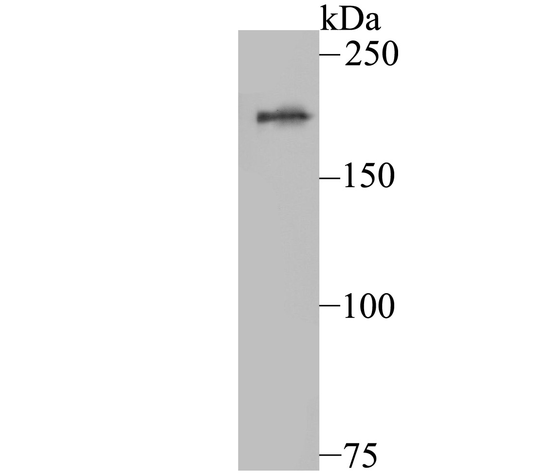
Western blot analysis of KDM5A on A549 cell using anti-KDM5A antibody at 1/500 dilution.ICC/IF
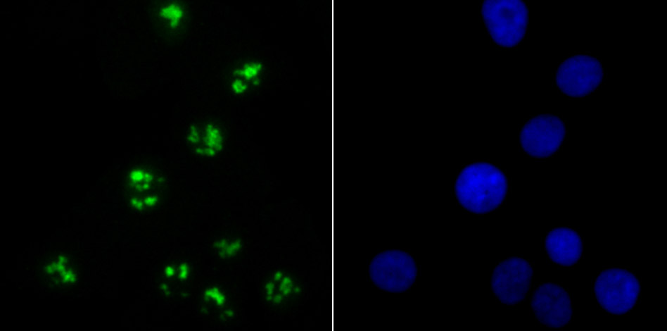
ICC staining KDM5A in A549 cells (green). The nuclear counter stain is DAPI (blue). Cells were fixed in paraformaldehyde, permeabilised with 0.25% Triton X100/PBS.ICC/IF
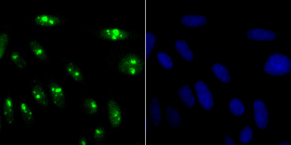
ICC staining KDM5A in SiHa cells (green). The nuclear counter stain is DAPI (blue). Cells were fixed in paraformaldehyde, permeabilised with 0.25% Triton X100/PBS.FC
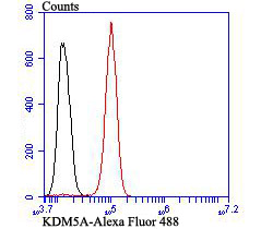
Flow cytometric analysis of SH-SY-5Y cells with KDM5A antibody at 1/100 dilution (red) compared with an unlabelled control (cells without incubation with primary antibody; black). Alexa Fluor 488-conjugated goat anti-rabbit IgG was used as the secondary antibody.WB
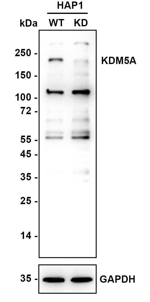
Western blot analysis of KDM5A on different lysates with Rabbit anti-KDM5A antibody at 1/2,000 dilution. Lane 1: HAP1-parental cell lysate, 10 µg/Lane, Lane 2: HAP1-KDM5A KD cell lysate, 10 µg/Lane, Exposure time: 180 seconds; 4-20% SDS-PAGE gel. Proteins were transferred to a PVDF membrane and blocked with 5% NFDM/TBST for 1 hour at room temperature. The primary antibody at 1/2,000 dilution was used in K1803 at 4℃ overnight. Goat Anti-Rabbit IgG - HRP Secondary Antibody at 1/50,000 dilution was used for 1 hour at room temperature.| Product Name | KDM5A Recombinant Rabbit Monoclonal Antibody |
|---|---|
| Antibody Type | Primary Antibodies |
| Product description | Histone demethylase that specifically demethylates 'Lys-4' of histone H3, thereby playing a central role in histone code. Does not demethylate histone H3 'Lys-9', H3 'Lys-27', H3 'Lys-36', H3 'Lys-79' or H4 'Lys-20'. Demethylates trimethylated and dimethylated but not monomethylated H3 'Lys-4'. Regulates specific gene transcription through DNA-binding on 5'-CCGCCC-3' motif. May stimulate transcription mediated by nuclear receptors. Involved in transcriptional regulation of Hox proteins during cell differentiation. May participate in transcriptional repression of cytokines such as CXCL12. Plays a role in the regulation of the circadian rhythm and in maintaining the normal periodicity of the circadian clock. In a histone demethylase-independent manner, acts as a coactivator of the CLOCK-ARNTL/BMAL1-mediated transcriptional activation of PER1/2 and other clock-controlled genes and increases histone acetylation at PER1/2 promoters by inhibiting the activity of HDAC1. Seems to act as a transcriptional corepressor for some genes such as MT1F and to favor the proliferation of cancer cells. |
| Immunogen | Recombinant protein within Human KDM5A aa 1,360-1,690 / 1,690. |
| Clonality | Monoclonal |
|---|---|
| Isotype | IgG |
| Host Species | Recombinant rabbit |
| Tested Applications | FCICC/IFWB |
| WB:1:1000-1:2000 ICC/IF:1:50-1:400 FC:1:50-1:100 |
|
| Species Reactivity | Human |
| Concentration | 1mg/ml |
| Purification | Protein A |
| Alternative Names | Histone demethylase JARID1A antibody JARID1A antibody Jumonji/ARID domain containing protein 1A antibody Jumonji/ARID domain-containing protein 1A antibody Kdm5a antibody KDM5A_HUMAN antibody Lysine-specific demethylase 5A antibody RBBP-2 antibody RBBP2 antibody RBP2 antibody Retinoblastoma binding protein 2 antibody Retinoblastoma-binding protein 2 antibody |
|---|---|
| Molecular Weight(MW) | 192 kDa(Observed band size: 200 kDa) |
| Cellular Localization | Nucleus. |
| SwissProt ID | P29375 |
|---|
WB
Western blot analysis of KDM5A on A549 cell using anti-KDM5A antibody at 1/500 dilution.
ICC/IF
ICC staining KDM5A in A549 cells (green). The nuclear counter stain is DAPI (blue). Cells were fixed in paraformaldehyde, permeabilised with 0.25% Triton X100/PBS.
ICC/IF
ICC staining KDM5A in SiHa cells (green). The nuclear counter stain is DAPI (blue). Cells were fixed in paraformaldehyde, permeabilised with 0.25% Triton X100/PBS.
FC
Flow cytometric analysis of SH-SY-5Y cells with KDM5A antibody at 1/100 dilution (red) compared with an unlabelled control (cells without incubation with primary antibody; black). Alexa Fluor 488-conjugated goat anti-rabbit IgG was used as the secondary antibody.
WB
Western blot analysis of KDM5A on different lysates with Rabbit anti-KDM5A antibody at 1/2,000 dilution. Lane 1: HAP1-parental cell lysate, 10 µg/Lane, Lane 2: HAP1-KDM5A KD cell lysate, 10 µg/Lane, Exposure time: 180 seconds; 4-20% SDS-PAGE gel. Proteins were transferred to a PVDF membrane and blocked with 5% NFDM/TBST for 1 hour at room temperature. The primary antibody at 1/2,000 dilution was used in K1803 at 4℃ overnight. Goat Anti-Rabbit IgG - HRP Secondary Antibody at 1/50,000 dilution was used for 1 hour at room temperature.| Application Notes | WB:1:1000-1:2000 ICC/IF:1:50-1:400 FC:1:50-1:100 |
|---|
| Form | Liquid |
|---|---|
| Storage Instructions | Store at +4℃ after thawing. Aliquot store at -20℃ or -80℃. Avoid repeated freeze / thaw cycles. |
| Storage Buffer | 1*TBS (pH7.4), 1%BSA, 40%Glycerol. Preservative: 0.05% Sodium Azide. |


