Application
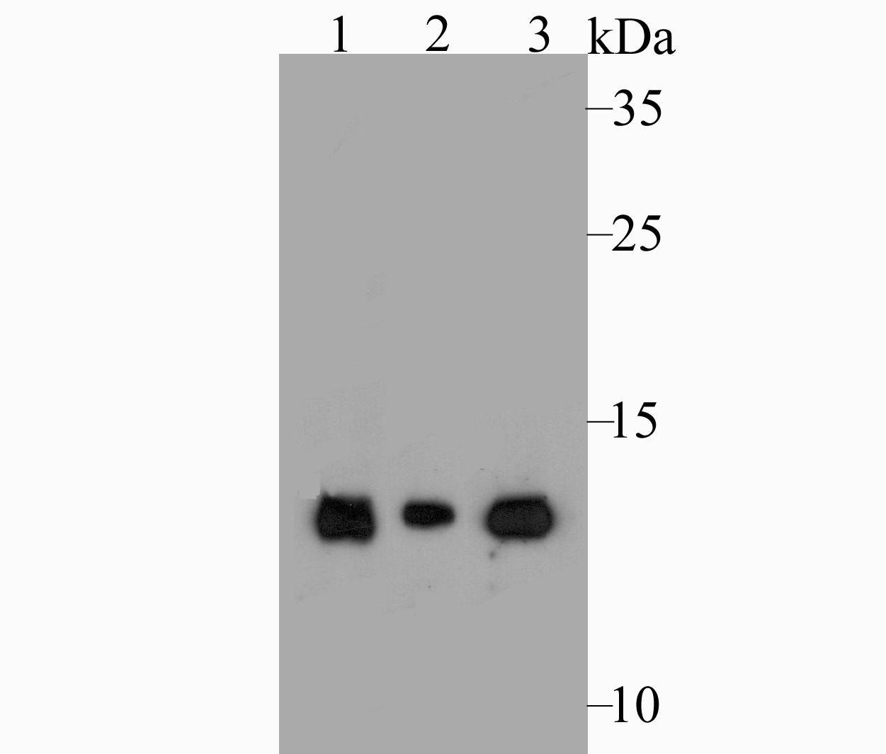
Fig1: Western blot analysis of Lysozyme on different lysates. Proteins were transferred to a PVDF membrane and blocked with 5% BSA in PBS for 1 hour at room temperature. The primary antibody was used at a 1:500 dilution in 5% BSA at room temperature for 2 hours. Goat Anti-Rabbit IgG - HRP Secondary Antibody (HA1001) at 1:5,000 dilution was used for 1 hour at room temperature. Positive control: Lane 1: HL-60 cell lysate Lane 2: Mouse kidney tissue lysate Lane 3: Rat kidney tissue lysateApplication
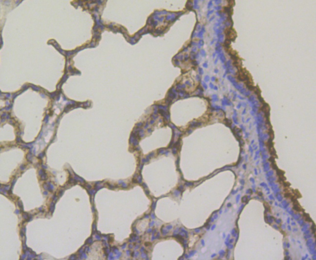
Fig2: Immunohistochemical analysis of paraffin-embedded rat lung tissue using anti-Lysozyme antibody. The section was pre-treated using heat mediated antigen retrieval with Tris-EDTA buffer (pH 8.0-8.4) for 20 minutes.The tissues were blocked in 5% BSA for 30 minutes at room temperature, washed with ddH2O and PBS, and then probed with the antibody (ER1803-88) at 1/200 dilution, for 30 minutes at room temperature and detected using an HRP conjugated compact polymer system. DAB was used as the chromogen. Counter stained with hematoxylin and mounted with DPX.Application
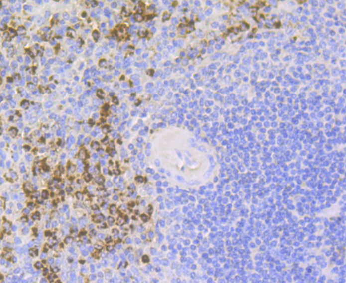
Fig3: Immunohistochemical analysis of paraffin-embedded human spleen tissue using anti-Lysozyme antibody. The section was pre-treated using heat mediated antigen retrieval with Tris-EDTA buffer (pH 8.0-8.4) for 20 minutes.The tissues were blocked in 5% BSA for 30 minutes at room temperature, washed with ddH2O and PBS, and then probed with the antibody (ER1803-88) at 1/200 dilution, for 30 minutes at room temperature and detected using an HRP conjugated compact polymer system. DAB was used as the chromogen. Counter stained with hematoxylin and mounted with DPX.Application
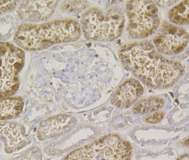
Fig4: Immunohistochemical analysis of paraffin-embedded human kidney tissue using anti-Lysozyme antibody. The section was pre-treated using heat mediated antigen retrieval with Tris-EDTA buffer (pH 8.0-8.4) for 20 minutes.The tissues were blocked in 5% BSA for 30 minutes at room temperature, washed with ddH2O and PBS, and then probed with the antibody (ER1803-88) at 1/200 dilution, for 30 minutes at room temperature and detected using an HRP conjugated compact polymer system. DAB was used as the chromogen. Counter stained with hematoxylin and mounted with DPX.Application
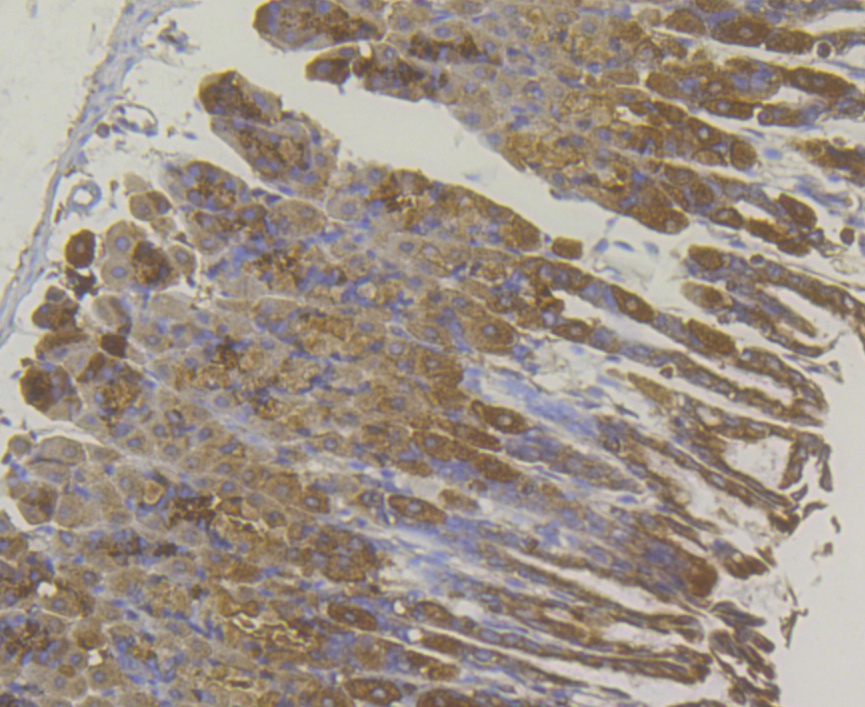
Fig5: Immunohistochemical analysis of paraffin-embedded mouse stomach tissue using anti-Lysozyme antibody. The section was pre-treated using heat mediated antigen retrieval with Tris-EDTA buffer (pH 8.0-8.4) for 20 minutes.The tissues were blocked in 5% BSA for 30 minutes at room temperature, washed with ddH2O and PBS, and then probed with the antibody (ER1803-88) at 1/50 dilution, for 30 minutes at room temperature and detected using an HRP conjugated compact polymer system. DAB was used as the chromogen. Counter stained with hematoxylin and mounted with DPX.| Product Name | Lysozyme |
|---|---|
| Antibody Type | Primary Antibodies |
| Antigen Alias | 1 4 beta N acetylmuramidase C antibody 1 antibody 4-beta-N-acetylmuramidase C antibody EC 3.2.1.17 antibody LYSC_HUMAN antibody Lysosyme antibody Lysozyme (renal amyloidosis) antibody Lysozyme C antibody Lysozyme C precursor antibody LYZ antibody LZM antibody Renal amyloidosis antibody |
| Product description | The origins of the lysozyme proteins date back an estimated 400 to 600 million years. Generally, lysozyme genes are relatively small, roughly 10 kilobases in length, and composed of four exons and three introns. Originally a bacteriolytic defensive agent, the function of this family of proteins adapted to serve a digestive function in its present forms. Lysozymes in tissues and body fluids are associated with the monocyte-macrophage system and enhance the activity of immunoagents. Lysozyme C belongs to the glycosyl hydrolase 22 family, and newly identified relatives of Lysozyme C appear to possess anti-HIV activity, as well as preserved bacteriolytic function against Micrococcus lysodeikticus. Lysozyme C is capable of both hydrolysis and transglycosylation and also a slight esterase activity. It acts rapidly on both peptide-substituted and unsubstituted peptidoglycan, and slowly on chitin oligosaccharides. Lysozyme C defects are a cause of amyloidosis VIII, also called familial visceral or Ostertag-type amyloidosis. |
| Immunogen | Recombinant protein within Human Lysozyme aa 19-148. |
| Clonality | Polyclonal |
|---|---|
| Isotype | IgG |
| Host Species | Rabbit |
| Tested Applications | WBIHC |
| WB:1:500-1:1,000: IHC:1:50-1:200: |
|
| Species Reactivity | HumanMouseRat |
| Concentration | 1mg/ml |
| Alternative Names | 1 4 beta N acetylmuramidase C antibody 1 antibody 4-beta-N-acetylmuramidase C antibody EC 3.2.1.17 antibody LYSC_HUMAN antibody Lysosyme antibody Lysozyme (renal amyloidosis) antibody Lysozyme C antibody Lysozyme C precursor antibody LYZ antibody LZM antibody Renal amyloidosis antibody |
|---|---|
| Molecular Weight(MW) | 17 kDa |
| Cellular Localization | Secreted. |
Application
Fig1: Western blot analysis of Lysozyme on different lysates. Proteins were transferred to a PVDF membrane and blocked with 5% BSA in PBS for 1 hour at room temperature. The primary antibody was used at a 1:500 dilution in 5% BSA at room temperature for 2 hours. Goat Anti-Rabbit IgG - HRP Secondary Antibody (HA1001) at 1:5,000 dilution was used for 1 hour at room temperature. Positive control: Lane 1: HL-60 cell lysate Lane 2: Mouse kidney tissue lysate Lane 3: Rat kidney tissue lysate
Application
Fig2: Immunohistochemical analysis of paraffin-embedded rat lung tissue using anti-Lysozyme antibody. The section was pre-treated using heat mediated antigen retrieval with Tris-EDTA buffer (pH 8.0-8.4) for 20 minutes.The tissues were blocked in 5% BSA for 30 minutes at room temperature, washed with ddH2O and PBS, and then probed with the antibody (ER1803-88) at 1/200 dilution, for 30 minutes at room temperature and detected using an HRP conjugated compact polymer system. DAB was used as the chromogen. Counter stained with hematoxylin and mounted with DPX.
Application
Fig3: Immunohistochemical analysis of paraffin-embedded human spleen tissue using anti-Lysozyme antibody. The section was pre-treated using heat mediated antigen retrieval with Tris-EDTA buffer (pH 8.0-8.4) for 20 minutes.The tissues were blocked in 5% BSA for 30 minutes at room temperature, washed with ddH2O and PBS, and then probed with the antibody (ER1803-88) at 1/200 dilution, for 30 minutes at room temperature and detected using an HRP conjugated compact polymer system. DAB was used as the chromogen. Counter stained with hematoxylin and mounted with DPX.
Application
Fig4: Immunohistochemical analysis of paraffin-embedded human kidney tissue using anti-Lysozyme antibody. The section was pre-treated using heat mediated antigen retrieval with Tris-EDTA buffer (pH 8.0-8.4) for 20 minutes.The tissues were blocked in 5% BSA for 30 minutes at room temperature, washed with ddH2O and PBS, and then probed with the antibody (ER1803-88) at 1/200 dilution, for 30 minutes at room temperature and detected using an HRP conjugated compact polymer system. DAB was used as the chromogen. Counter stained with hematoxylin and mounted with DPX.
Application
Fig5: Immunohistochemical analysis of paraffin-embedded mouse stomach tissue using anti-Lysozyme antibody. The section was pre-treated using heat mediated antigen retrieval with Tris-EDTA buffer (pH 8.0-8.4) for 20 minutes.The tissues were blocked in 5% BSA for 30 minutes at room temperature, washed with ddH2O and PBS, and then probed with the antibody (ER1803-88) at 1/50 dilution, for 30 minutes at room temperature and detected using an HRP conjugated compact polymer system. DAB was used as the chromogen. Counter stained with hematoxylin and mounted with DPX.| Positive Control | HL-60, mouse kidney tissue, rat kidney tissue, rat lung tissue, human spleen tissue, human kidney tissue, mouse stomach tissue. |
|---|---|
| Application Notes | WB:1:500-1:1,000: IHC:1:50-1:200: |
| Form | Liquid |
|---|---|
| Storage Instructions | Store at +4℃ after thawing. Aliquot store at -20℃. Avoid repeated freeze / thaw cycles. |
| Storage Buffer | 1*TBS (pH7.4), 1%BSA, 50%Glycerol. Preservative: 0.05% Sodium Azide. |


