Application
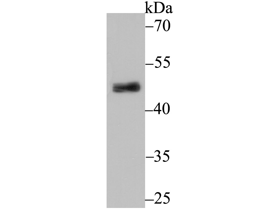
Fig1: Western blot analysis of PDK2 on rat heart tissue lysate using anti-PDK2 antibody at 1/500 dilution.Application
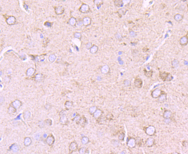
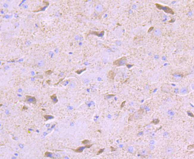
Fig2: Immunohistochemical analysis of paraffin-embedded rat brain tissue using anti-PDK2 antibody. Counter stained with hematoxylin.Application
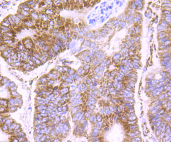
Fig3: Immunohistochemical analysis of paraffin-embedded human colon cancer tissue using anti-PDK2 antibody. Counter stained with hematoxylin.Application
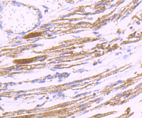
Fig4: Immunohistochemical analysis of paraffin-embedded human fetal skeletal muscle tissue using anti-PDK2 antibody. Counter stained with hematoxylin.Application
Fig5: Immunohistochemical analysis of paraffin-embedded mouse cerebellum tissue using anti-PDK2 antibody. Counter stained with hematoxylin.Application
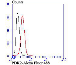
Fig6: Flow cytometric analysis of SH-SY-5Y cells with PDK2 antibody at 1/100 dilution (red) compared with an unlabelled control (cells without incubation with primary antibody; black). Alexa Fluor 488-conjugated goat anti-rabbit IgG was used as the secondary antibody.| Product Name | PDK2 [JB66-93] |
|---|---|
| Antibody Type | Primary Antibodies |
| Antigen Alias | [Pyruvate dehydrogenase [lipoamide]] kinase isozyme 2 antibody mitochondrial antibody PDHK2 antibody PDK2 antibody PDK2_HUMAN antibody Pyruvate dehydrogenase kinase isoform 2 antibody Pyruvate dehydrogenase kinase, isozyme 2 antibody Pyruvate dehydrogenase lipoamide kinase isozyme 2, mitochondrial antibody |
| Product description | Pyruvate dehydrogenase kinase family members (PDK1, 2, 3, 4) are serine kinases that catalyze the phosphorylation of the E1α subunit of the pyruvate dehydrogenase complex (PDC). PDC activity is controlled through phosphorylation and dephosphorylation of the E1α subunit, which leads to inactivation and reactivation, respectively. The core of PDC is composed of sixty dihydrolypoyl acetyltransferase (E2) subunits that bind directly to PDK2 and enhance PDK2 kinase activity. Upregulation of PDK isoenzymes occurs during starvation conditions, rerouting acetyl-CoA generation by facilitating fatty acid oxidation. PDKs contain five conserved regions and are mechanistically similar to bacterial His-kinases, in that both require Histidine residues for activity. In mammals, transcripts for PDK2 are ubiquitously expressed with high levels in heart and skeletal muscle and decreased levels in spleen and lung. |
| Immunogen | Recombinant protein |
| Clonality | Monoclonal |
|---|---|
| Isotype | IgG |
| Host Species | Recombinant rabbit |
| Tested Applications | WBIHCFCIP |
| WB:1:500 IHC:1:50-1:200 FC:1:50-1:100 |
|
| Species Reactivity | HumanMouseRat |
| Concentration | 1mg/ml |
| Alternative Names | [Pyruvate dehydrogenase [lipoamide]] kinase isozyme 2 antibody
mitochondrial antibody
PDHK2 antibody
PDK2 antibody
PDK2_HUMAN antibody
Pyruvate dehydrogenase kinase isoform 2 antibody
Pyruvate dehydrogenase kinase isozyme 2 antibody Pyruvate dehydrogenase lipoamide kinase isozyme 2 mitochondrial antibody |
|---|---|
| Molecular Weight(MW) | 46 kDa |
| Cellular Localization | Mitochondrion. |
| SwissProt ID | Q15119 |
|---|
Application
Fig1: Western blot analysis of PDK2 on rat heart tissue lysate using anti-PDK2 antibody at 1/500 dilution.
Application
Fig2: Immunohistochemical analysis of paraffin-embedded rat brain tissue using anti-PDK2 antibody. Counter stained with hematoxylin.
Application
Fig3: Immunohistochemical analysis of paraffin-embedded human colon cancer tissue using anti-PDK2 antibody. Counter stained with hematoxylin.
Application
Fig4: Immunohistochemical analysis of paraffin-embedded human fetal skeletal muscle tissue using anti-PDK2 antibody. Counter stained with hematoxylin.
Application
Fig5: Immunohistochemical analysis of paraffin-embedded mouse cerebellum tissue using anti-PDK2 antibody. Counter stained with hematoxylin.
Application
Fig6: Flow cytometric analysis of SH-SY-5Y cells with PDK2 antibody at 1/100 dilution (red) compared with an unlabelled control (cells without incubation with primary antibody; black). Alexa Fluor 488-conjugated goat anti-rabbit IgG was used as the secondary antibody.| Positive Control | Rat heart tissue lysate, rat brain tissue, human colon cancer tissue, human fetal skeletal muscle tissue, mouse cerebellum tissue, SH-SY-5Y. |
|---|---|
| Application Notes | WB:1:500 IHC:1:50-1:200 FC:1:50-1:100 |
| Form | Liquid |
|---|---|
| Storage Instructions | Store at +4℃ after thawing. Aliquot store at -20℃ or -80℃. Avoid repeated freeze / thaw cycles. |
| Storage Buffer | 1*TBS (pH7.4), 1%BSA, 40%Glycerol. Preservative: 0.05% Sodium Azide. |


