Application
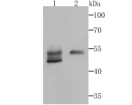
Fig1: Western blot analysis of Biglycan on mouse heart and human skin tissue lysates using anti-Biglycan antibody at 1/500 dilution.Application
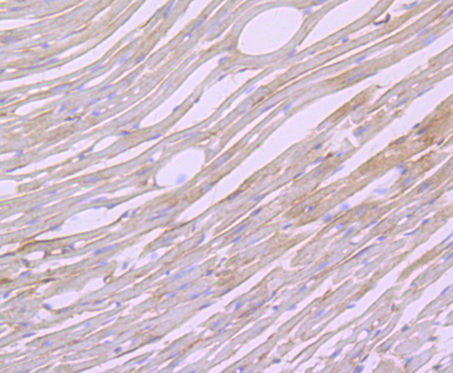
Fig2: Immunohistochemical analysis of paraffin-embedded mouse heart tissue using anti-Biglycan antibody. Counter stained with hematoxylin.Application
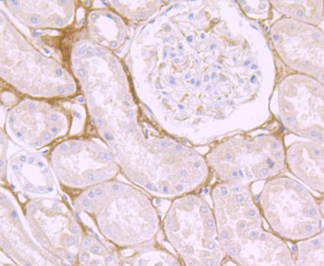
Fig3: Immunohistochemical analysis of paraffin-embedded human kidney tissue using anti-Biglycan antibody. Counter stained with hematoxylin.Application
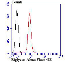
Fig4: Flow cytometric analysis of HepG2 cells with Biglycan antibody at 1/100 dilution (red) compared with an unlabelled control (cells without incubation with primary antibody; black). Alexa Fluor 488-conjugated goat anti-rabbit IgG was used as the secondary antibody.| Product Name | Biglycan [JB71-31] |
|---|---|
| Antibody Type | Primary Antibodies |
| Antigen Alias | BGN antibody Biglycan antibody Biglycan proteoglycan antibody Bone/cartilage proteoglycan I antibody Dermatan sulphate proteoglycan I antibody DSPG1 antibody PG S1 antibody PG-S1 antibody PGI antibody PGS1_HUMAN antibody SEMDX antibody SLRR1A antibody Small leucine rich protein 1A antibody |
| Product description | Biglycan, a class I small leucine rich proteoglycan (SLRP) present in the extracellular matrix, influences bone cell differentiation and proliferation. Biglycan contains two chondroitin sulfate glucosaminoglycan (GAG) chains attached near its amino terminus, whereas a closely related SLRP, decorin, contains only one. Biglycan deficient specimens possess diminished capacity to produce bone cells precursors, a lessened response to TGFβ, reduced collagen synthesis, and increased apoptosis. Patients with rheumatoid arthritis express increased immunity to biglycan whereas osteoarthritis patients do not, suggesting that higher immunity to SLRPs may play a role in the pathogenesis of inflammatory rheumatic diseases. |
| Immunogen | Recombinant protein |
| Clonality | Monoclonal |
|---|---|
| Isotype | IgG |
| Host Species | Recombinant rabbit |
| Tested Applications | WBIHCFCIP |
| WB:1:500-1:1000 IHC:1:50-1:200 FC:1:50-1:100 |
|
| Species Reactivity | HumanMouse |
| Concentration | 1mg/ml |
| Alternative Names | BGN antibody Biglycan antibody Biglycan proteoglycan antibody Bone/cartilage proteoglycan I antibody Dermatan sulphate proteoglycan I antibody DSPG1 antibody PG S1 antibody PG-S1 antibody PGI antibody PGS1_HUMAN antibody SEMDX antibody SLRR1A antibody Small leucine rich protein 1A antibody |
|---|---|
| Molecular Weight(MW) | 42 kDa |
| Cellular Localization | Extracellular matrix. Secreted. |
Application
Fig1: Western blot analysis of Biglycan on mouse heart and human skin tissue lysates using anti-Biglycan antibody at 1/500 dilution.
Application
Fig2: Immunohistochemical analysis of paraffin-embedded mouse heart tissue using anti-Biglycan antibody. Counter stained with hematoxylin.
Application
Fig3: Immunohistochemical analysis of paraffin-embedded human kidney tissue using anti-Biglycan antibody. Counter stained with hematoxylin.
Application
Fig4: Flow cytometric analysis of HepG2 cells with Biglycan antibody at 1/100 dilution (red) compared with an unlabelled control (cells without incubation with primary antibody; black). Alexa Fluor 488-conjugated goat anti-rabbit IgG was used as the secondary antibody.| Positive Control | HepG2, human kidney tissue, mouse heart tissue, human skin tissue lysates. |
|---|---|
| Application Notes | WB:1:500-1:1000 IHC:1:50-1:200 FC:1:50-1:100 |
| Form | Liquid |
|---|---|
| Storage Instructions | Store at +4℃ after thawing. Aliquot store at -20℃ or -80℃. Avoid repeated freeze / thaw cycles. |
| Storage Buffer | 1*TBS (pH7.4), 1%BSA, 40%Glycerol. Preservative: 0.05% Sodium Azide. |


