WB
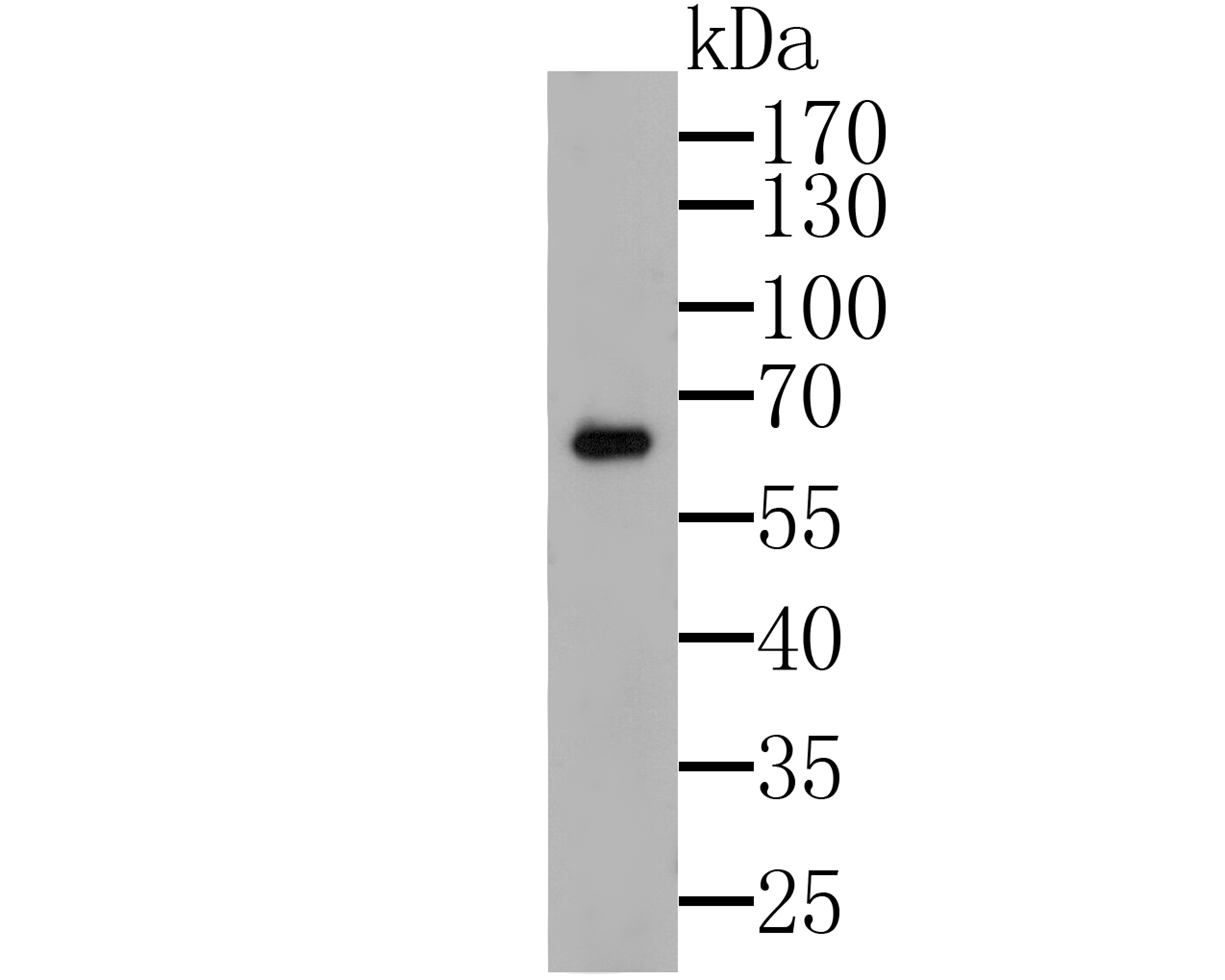
Western blot analysis of G-protein coupled receptor 30 on Lovo cell lysate. Proteins were transferred to a PVDF membrane and blocked with 5% BSA in PBS for 1 hour at room temperature. The primary antibody was used at a 1:500 dilution in 5% BSA at room temperature for 2 hours. Goat Anti-Rabbit IgG - HRP Secondary Antibody (HA1001) at 1:5,000 dilution was used for 1 hour at room temperature.ICC/IF
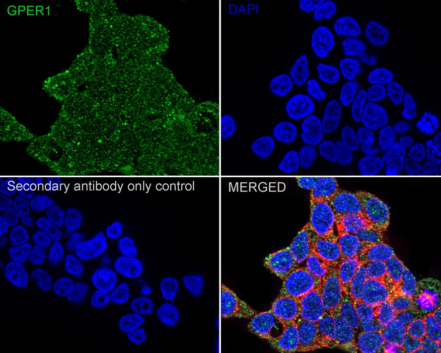
Immunocytochemistry analysis of LoVo cells labeling G-protein coupled receptor 30 with Rabbit anti-G-protein coupled receptor 30 antibody at 1/100 dilution. Cells were fixed in 4% paraformaldehyde for 15 minutes at room temperature, permeabilized with 0.1% Triton X-100 in PBS for 15 minutes at room temperature, then blocked with 1% BSA in 10% negative goat serum for 1 hour at room temperature. Cells were then incubated with Rabbit anti-G-protein coupled receptor 30 antibody at 1/100 dilution in 1% BSA in PBST overnight at 4 ℃. Goat Anti-Rabbit IgG H&L (488) was used as the secondary antibody at 1/1,000 dilution. PBS instead of the primary antibody was used as the secondary antibody only control. Nuclear DNA was labelled in blue with DAPI. Beta tubulin (red) was stained at 1/100 dilution overnight at +4℃. Goat Anti-Mouse IgG H&L (594) was used as the secondary antibody at 1/1,000 dilution.ICC/IF
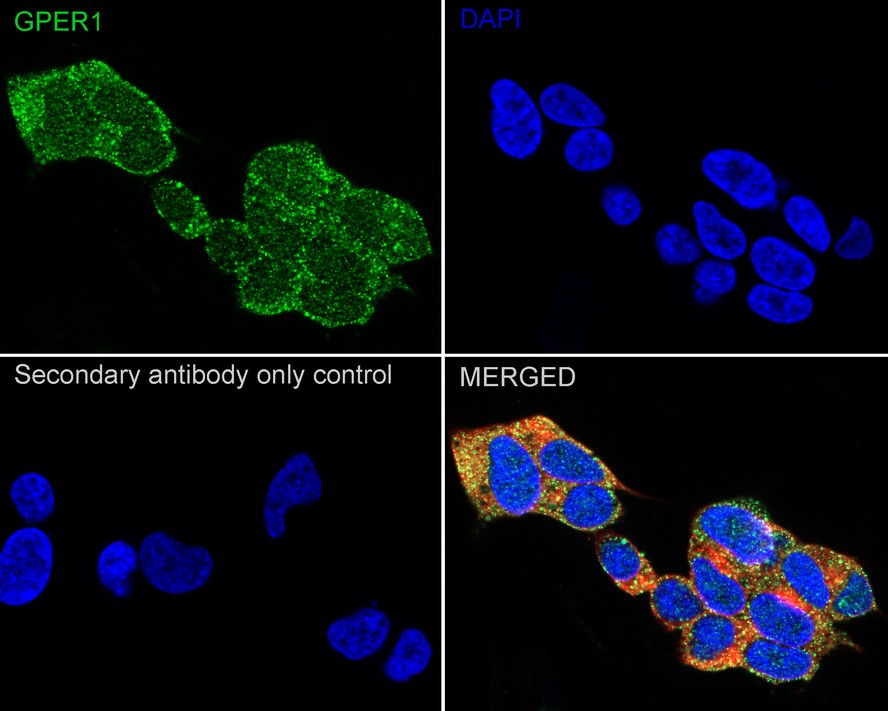
Immunocytochemistry analysis of SH-SY5Y cells labeling G-protein coupled receptor 30 with Rabbit anti-G-protein coupled receptor 30 antibody at 1/100 dilution. Cells were fixed in 4% paraformaldehyde for 15 minutes at room temperature, permeabilized with 0.1% Triton X-100 in PBS for 15 minutes at room temperature, then blocked with 1% BSA in 10% negative goat serum for 1 hour at room temperature. Cells were then incubated with Rabbit anti-G-protein coupled receptor 30 antibody at 1/100 dilution in 1% BSA in PBST overnight at 4 ℃. Goat Anti-Rabbit IgG H&L (488) was used as the secondary antibody at 1/1,000 dilution. PBS instead of the primary antibody was used as the secondary antibody only control. Nuclear DNA was labelled in blue with DAPI. Beta tubulin (red) was stained at 1/100 dilution overnight at +4℃. Goat Anti-Mouse IgG H&L (594) was used as the secondary antibody at 1/1,000 dilution.IHC
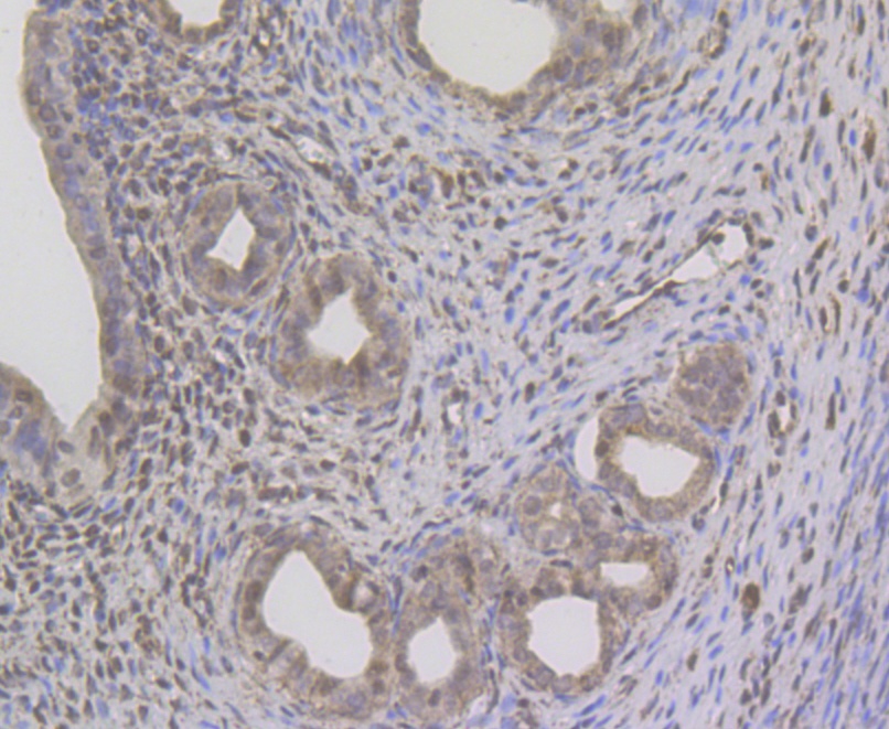
Immunohistochemical analysis of paraffin-embedded rat uterus tissue using anti-G-protein coupled receptor 30 antibody. The section was pre-treated using heat mediated antigen retrieval with Tris-EDTA buffer (pH 8.0-8.4) for 20 minutes.The tissues were blocked in 5% BSA for 30 minutes at room temperature, washed with ddH2O and PBS, and then probed with the antibody (ER1803-90) at 1/200 dilution, for 30 minutes at room temperature and detected using an HRP conjugated compact polymer system. DAB was used as the chrogen. Counter stained with hematoxylin and mounted with DPX.IHC
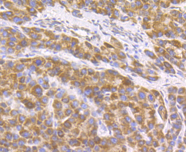
Immunohistochemical analysis of paraffin-embedded human liver cancer tissue using anti-G-protein coupled receptor 30 antibody. The section was pre-treated using heat mediated antigen retrieval with Tris-EDTA buffer (pH 8.0-8.4) for 20 minutes.The tissues were blocked in 5% BSA for 30 minutes at room temperature, washed with ddH2O and PBS, and then probed with the antibody (ER1803-90) at 1/200 dilution, for 30 minutes at room temperature and detected using an HRP conjugated compact polymer system. DAB was used as the chrogen. Counter stained with hematoxylin and mounted with DPX.IHC
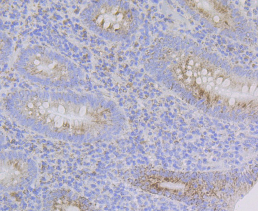
Immunohistochemical analysis of paraffin-embedded human appendix tissue using anti-G-protein coupled receptor 30 antibody. The section was pre-treated using heat mediated antigen retrieval with Tris-EDTA buffer (pH 8.0-8.4) for 20 minutes.The tissues were blocked in 5% BSA for 30 minutes at room temperature, washed with ddH2O and PBS, and then probed with the antibody (ER1803-90) at 1/200 dilution, for 30 minutes at room temperature and detected using an HRP conjugated compact polymer system. DAB was used as the chrogen. Counter stained with hematoxylin and mounted with DPX.IHC
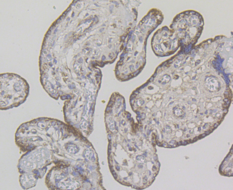
Immunohistochemical analysis of paraffin-embedded human placenta tissue using anti-G-protein coupled receptor 30 antibody. The section was pre-treated using heat mediated antigen retrieval with Tris-EDTA buffer (pH 8.0-8.4) for 20 minutes.The tissues were blocked in 5% BSA for 30 minutes at room temperature, washed with ddH2O and PBS, and then probed with the antibody (ER1803-90) at 1/200 dilution, for 30 minutes at room temperature and detected using an HRP conjugated compact polymer system. DAB was used as the chrogen. Counter stained with hematoxylin and mounted with DPX.IHC
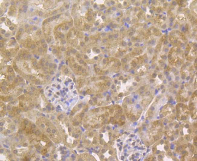
Immunohistochemical analysis of paraffin-embedded mouse kidney tissue using anti-G-protein coupled receptor 30 antibody. The section was pre-treated using heat mediated antigen retrieval with Tris-EDTA buffer (pH 8.0-8.4) for 20 minutes.The tissues were blocked in 5% BSA for 30 minutes at room temperature, washed with ddH2O and PBS, and then probed with the antibody (ER1803-90) at 1/200 dilution, for 30 minutes at room temperature and detected using an HRP conjugated compact polymer system. DAB was used as the chrogen. Counter stained with hematoxylin and mounted with DPX.FC
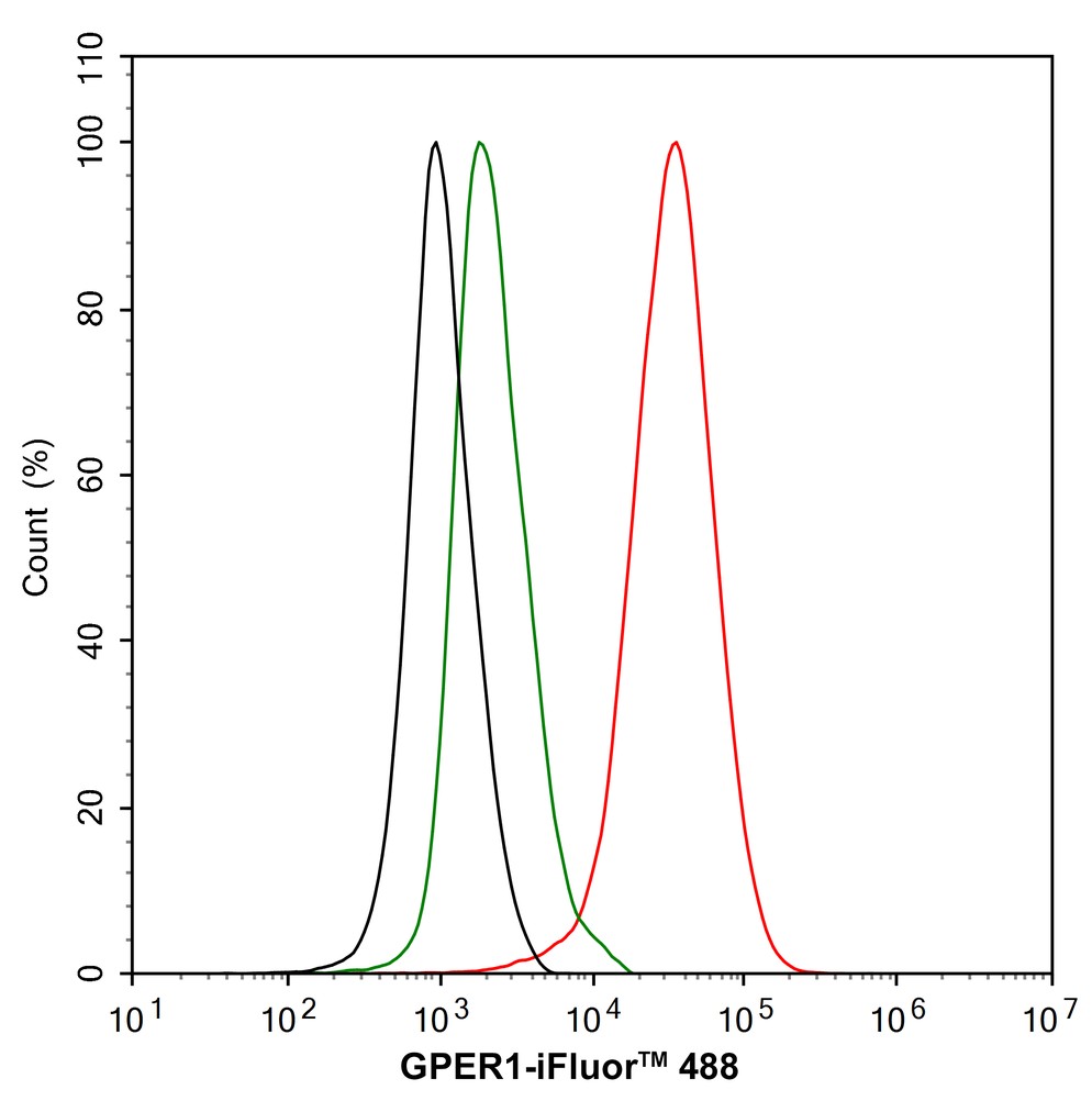
Flow cytometric analysis of SH-SY5Y cells labeling G-protein coupled receptor 30. Cells were fixed and permeabilized. Then stained with the primary antibody (1/1,000) (red) compared with Rabbit IgG Isotype Control (green). After incubation of the primary antibody at +4℃ for an hour, the cells were stained with a 488 conjugate-Goat anti-Rabbit IgG Secondary antibody at 1/1,000 dilution for 30 minutes at +4℃. Unlabelled sample was used as a control (cells without incubation with primary antibody; black).| Product Name | G-protein coupled receptor 30 Rabbit Polyclonal Antibody |
|---|---|
| Antibody Type | Primary Antibodies |
| Product description | G-protein coupled estrogen receptor that binds to 17-beta-estradiol (E2) with high affinity, leading to rapid and transient activation of numerous intracellular signaling pathways. Stimulates cAMP production, calcium mobilization and tyrosine kinase Src inducing the release of heparin-bound epidermal growth factor (HB-EGF) and subsequent transactivation of the epidermal growth factor receptor (EGFR), activating downstream signaling pathways such as PI3K/Akt and ERK/MAPK. Mediates pleiotropic functions among others in the cardiovascular, endocrine, reproductive, immune and central nervous systems. Triggers mitochondrial apoptosis during pachytene spermatocyte differentiation. Stimulates uterine epithelial cell proliferation. Enhances uterine contractility in response to oxytocin. Contributes to thymic atrophy by inducing apoptosis. Increases firing activity and intracellular calcium oscillations in luteinizing hormone-releasing hormone (LHRH) neurons. Inhibits early osteoblast proliferation at growth plate during skeletal development. Inhibits mature adipocyte differentiation and lipid accumulation. Stimulates cancer-associated fibroblast (CAF) proliferation by a rapid genomic response through the EGFR/ERK transduction pathway. Associated with EGFR, may act as a transcription factor activating growth regulatory genes (c-fos, cyclin D1). Promotes integrin alpha-5/beta-1 and fibronectin (FN) matrix assembly in breast cancer cells. |
| Immunogen | Recombinant protein within Human G-protein coupled receptor 30 aa 236-375 / 375. |
| Clonality | Polyclonal |
|---|---|
| Isotype | IgG |
| Host Species | Rabbit |
| Tested Applications | FCICC/IFIHCWB |
| WWB:1:500-1:1000 ICC/IF:1:100 IHC:1:50-1:200 FC:1:1000 |
|
| Species Reactivity | HumanMouseRat |
| Concentration | 1mg/ml |
| Purification | Affinity purified |
| Alternative Names | CEPR antibody Chemoattractant receptor-like 2 antibody Chemokine receptor-like 2 antibody CMKRL2 antibody Constitutively expressed peptide like receptor antibody DRY12 antibody FEG 1 antibody FEG-1 antibody Flow-induced endothelial G protein-coupled receptor antibody Flow-induced endothelial G-protein coupled receptor 1 antibody G protein-coupled receptor 30 antibody G-protein coupled estrogen receptor 1 antibody G-protein coupled receptor 30 antibody GPCR-BR antibody Gper antibody GPER_HUMAN antibody GPER1 antibody GPR30 antibody IL8-related receptor DRY12 antibody Lergu antibody LERGU2 antibody leucine rich protein in GPR30 3'UTR antibody LYGPR antibody Lymphocyte-derived G-protein coupled receptor antibody Membrane estrogen receptor antibody mER antibody MGC99678 antibody |
|---|---|
| Molecular Weight(MW) | Predicted band size: 42 kDa |
| Cellular Localization | Mitochondrion,Nucleus,Endosome,Golgi apparatus,Endoplasmic reticulum,Cytoskeleton. |
WB
Western blot analysis of G-protein coupled receptor 30 on Lovo cell lysate. Proteins were transferred to a PVDF membrane and blocked with 5% BSA in PBS for 1 hour at room temperature. The primary antibody was used at a 1:500 dilution in 5% BSA at room temperature for 2 hours. Goat Anti-Rabbit IgG - HRP Secondary Antibody (HA1001) at 1:5,000 dilution was used for 1 hour at room temperature.
ICC/IF
Immunocytochemistry analysis of LoVo cells labeling G-protein coupled receptor 30 with Rabbit anti-G-protein coupled receptor 30 antibody at 1/100 dilution. Cells were fixed in 4% paraformaldehyde for 15 minutes at room temperature, permeabilized with 0.1% Triton X-100 in PBS for 15 minutes at room temperature, then blocked with 1% BSA in 10% negative goat serum for 1 hour at room temperature. Cells were then incubated with Rabbit anti-G-protein coupled receptor 30 antibody at 1/100 dilution in 1% BSA in PBST overnight at 4 ℃. Goat Anti-Rabbit IgG H&L (488) was used as the secondary antibody at 1/1,000 dilution. PBS instead of the primary antibody was used as the secondary antibody only control. Nuclear DNA was labelled in blue with DAPI. Beta tubulin (red) was stained at 1/100 dilution overnight at +4℃. Goat Anti-Mouse IgG H&L (594) was used as the secondary antibody at 1/1,000 dilution.
ICC/IF
Immunocytochemistry analysis of SH-SY5Y cells labeling G-protein coupled receptor 30 with Rabbit anti-G-protein coupled receptor 30 antibody at 1/100 dilution. Cells were fixed in 4% paraformaldehyde for 15 minutes at room temperature, permeabilized with 0.1% Triton X-100 in PBS for 15 minutes at room temperature, then blocked with 1% BSA in 10% negative goat serum for 1 hour at room temperature. Cells were then incubated with Rabbit anti-G-protein coupled receptor 30 antibody at 1/100 dilution in 1% BSA in PBST overnight at 4 ℃. Goat Anti-Rabbit IgG H&L (488) was used as the secondary antibody at 1/1,000 dilution. PBS instead of the primary antibody was used as the secondary antibody only control. Nuclear DNA was labelled in blue with DAPI. Beta tubulin (red) was stained at 1/100 dilution overnight at +4℃. Goat Anti-Mouse IgG H&L (594) was used as the secondary antibody at 1/1,000 dilution.
IHC
Immunohistochemical analysis of paraffin-embedded rat uterus tissue using anti-G-protein coupled receptor 30 antibody. The section was pre-treated using heat mediated antigen retrieval with Tris-EDTA buffer (pH 8.0-8.4) for 20 minutes.The tissues were blocked in 5% BSA for 30 minutes at room temperature, washed with ddH2O and PBS, and then probed with the antibody (ER1803-90) at 1/200 dilution, for 30 minutes at room temperature and detected using an HRP conjugated compact polymer system. DAB was used as the chrogen. Counter stained with hematoxylin and mounted with DPX.
IHC
Immunohistochemical analysis of paraffin-embedded human liver cancer tissue using anti-G-protein coupled receptor 30 antibody. The section was pre-treated using heat mediated antigen retrieval with Tris-EDTA buffer (pH 8.0-8.4) for 20 minutes.The tissues were blocked in 5% BSA for 30 minutes at room temperature, washed with ddH2O and PBS, and then probed with the antibody (ER1803-90) at 1/200 dilution, for 30 minutes at room temperature and detected using an HRP conjugated compact polymer system. DAB was used as the chrogen. Counter stained with hematoxylin and mounted with DPX.
IHC
Immunohistochemical analysis of paraffin-embedded human appendix tissue using anti-G-protein coupled receptor 30 antibody. The section was pre-treated using heat mediated antigen retrieval with Tris-EDTA buffer (pH 8.0-8.4) for 20 minutes.The tissues were blocked in 5% BSA for 30 minutes at room temperature, washed with ddH2O and PBS, and then probed with the antibody (ER1803-90) at 1/200 dilution, for 30 minutes at room temperature and detected using an HRP conjugated compact polymer system. DAB was used as the chrogen. Counter stained with hematoxylin and mounted with DPX.
IHC
Immunohistochemical analysis of paraffin-embedded human placenta tissue using anti-G-protein coupled receptor 30 antibody. The section was pre-treated using heat mediated antigen retrieval with Tris-EDTA buffer (pH 8.0-8.4) for 20 minutes.The tissues were blocked in 5% BSA for 30 minutes at room temperature, washed with ddH2O and PBS, and then probed with the antibody (ER1803-90) at 1/200 dilution, for 30 minutes at room temperature and detected using an HRP conjugated compact polymer system. DAB was used as the chrogen. Counter stained with hematoxylin and mounted with DPX.
IHC
Immunohistochemical analysis of paraffin-embedded mouse kidney tissue using anti-G-protein coupled receptor 30 antibody. The section was pre-treated using heat mediated antigen retrieval with Tris-EDTA buffer (pH 8.0-8.4) for 20 minutes.The tissues were blocked in 5% BSA for 30 minutes at room temperature, washed with ddH2O and PBS, and then probed with the antibody (ER1803-90) at 1/200 dilution, for 30 minutes at room temperature and detected using an HRP conjugated compact polymer system. DAB was used as the chrogen. Counter stained with hematoxylin and mounted with DPX.
FC
Flow cytometric analysis of SH-SY5Y cells labeling G-protein coupled receptor 30. Cells were fixed and permeabilized. Then stained with the primary antibody (1/1,000) (red) compared with Rabbit IgG Isotype Control (green). After incubation of the primary antibody at +4℃ for an hour, the cells were stained with a 488 conjugate-Goat anti-Rabbit IgG Secondary antibody at 1/1,000 dilution for 30 minutes at +4℃. Unlabelled sample was used as a control (cells without incubation with primary antibody; black).| Application Notes | WWB:1:500-1:1000 ICC/IF:1:100 IHC:1:50-1:200 FC:1:1000 |
|---|
| Form | Liquid |
|---|---|
| Storage Instructions | Store at +4℃ after thawing. Aliquot store at -20℃. Avoid repeated freeze / thaw cycles. |
| Storage Buffer | 1*TBS (pH7.4), 1%BSA, 50%Glycerol. Preservative: 0.05% Sodium Azide. |


