Application
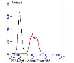
Fig4: Flow cytometric analysis of K562 cells with PU.1/Spi1 antibody at 1/100 dilution (red) compared with an unlabelled control (cells without incubation with primary antibody; black). Alexa Fluor 488-conjugated goat anti rabbit IgG was used as the secondary antibody.Application
Fig4: Flow cytometric analysis of K562 cells with PU.1/Spi1 antibody at 1/100 dilution (red) compared with an unlabelled control (cells without incubation with primary antibody; black). Alexa Fluor 488-conjugated goat anti rabbit IgG was used as the secondary antibody.Application
Fig4: Flow cytometric analysis of K562 cells with PU.1/Spi1 antibody at 1/100 dilution (red) compared with an unlabelled control (cells without incubation with primary antibody; black). Alexa Fluor 488-conjugated goat anti rabbit IgG was used as the secondary antibody.Application
Fig4: Flow cytometric analysis of K562 cells with PU.1/Spi1 antibody at 1/100 dilution (red) compared with an unlabelled control (cells without incubation with primary antibody; black). Alexa Fluor 488-conjugated goat anti rabbit IgG was used as the secondary antibody.| Product Name | PU.1/Spi1 [JB72-35] |
|---|---|
| Antibody Type | Primary Antibodies |
| Antigen Alias | transcription factor spi1 antibody 31 kDa Transforming Protein antibody 31 kDa-transforming protein antibody cb1086 antibody Hematopoietic transcription factor PU.1 antibody OF antibody oncogene spi1 antibody PU.1 antibody SFFV virus-induced murine erythroleukemia oncogene, mouse, homolog of antibody SFPI1 antibody si:by184l24.2 antibody SPI 1 antibody SPI 1 proto oncogene antibody SPI A antibody Spi1 antibody SPI1_HUMAN antibody Spleen focus forming virus (SFFV) proviral integration oncogene spi1 antibody Spleen focus forming virus proviral integration oncogene spi1 antibody Transcription factor PU.1 antibody |
| Product description | The Ets transcription factor family (Ets-1, Ets-2, Erg-1-3, Elk-1, Elf-1, Elf-5, NERF, PU.1, PEA3, ERM, FEV, ER8l, Fli-1, TEL, Spi-B, ESE-1, ESE-3A, Net, ABT1 and ERF) are DNA-binding proteins that influence lymphoid development and activity. The Ets family monomeric proteins bind the consensus DNA site GGA(A/T) through a unique winged helix-turn-helix motif known as the Ets domain. PU.1 (Spi-1/Spi-A), Spi-B and Spi-C are closely related Ets family members which share a conserved divergent sequence within the Ets domain that enables their binding to the non-canonical AGAA sites. PU.1 transactivates a large number of B cell genes, such as those encoding CD72, CD20 and Btk, and Spi-B enhances expression of many of these same target genes. PU.1 is expressed in a wide variety of hematopoetic cells, including B cells, early T-cells, megakaryocytes, granulocytes, mast cells, immature erythrocytes and myeloid cells. Alternatively, Spi-B expression is limited to B cells and immature T cells, where expression accumulates through T-lineage commitment and then is dramatically absent following the beta-selection checkpoint. |
| Immunogen | Recombinant protein |
| Clonality | Monoclonal |
|---|---|
| Isotype | IgG |
| Host Species | Recombinant rabbit |
| Tested Applications | WBIHCFC |
| WB:1:500-1:1,000 IHC:1:50-1:200 FC:1:50-1:100 |
|
| Species Reactivity | Human |
| Concentration | 1mg/ml |
| Alternative Names | transcription factor spi1 antibody
31 kDa Transforming Protein antibody
31 kDa-transforming protein antibody
cb1086 antibody
Hematopoietic transcription factor PU.1 antibody
OF antibody
oncogene spi1 antibody
PU.1 antibody
SFFV virus-induced murine erythroleukemia oncogene mouse homolog of antibody SFPI1 antibody si:by184l24.2 antibody SPI 1 antibody SPI 1 proto oncogene antibody SPI A antibody Spi1 antibody SPI1_HUMAN antibody Spleen focus forming virus (SFFV) proviral integration oncogene spi1 antibody Spleen focus forming virus proviral integration oncogene spi1 antibody Transcription factor PU.1 antibody |
|---|---|
| Molecular Weight(MW) | 31 kDa |
| Cellular Localization | Cytosol. |
| SwissProt ID | P17947 |
|---|
Application
Fig4: Flow cytometric analysis of K562 cells with PU.1/Spi1 antibody at 1/100 dilution (red) compared with an unlabelled control (cells without incubation with primary antibody; black). Alexa Fluor 488-conjugated goat anti rabbit IgG was used as the secondary antibody.
Application
Fig4: Flow cytometric analysis of K562 cells with PU.1/Spi1 antibody at 1/100 dilution (red) compared with an unlabelled control (cells without incubation with primary antibody; black). Alexa Fluor 488-conjugated goat anti rabbit IgG was used as the secondary antibody.
Application
Fig4: Flow cytometric analysis of K562 cells with PU.1/Spi1 antibody at 1/100 dilution (red) compared with an unlabelled control (cells without incubation with primary antibody; black). Alexa Fluor 488-conjugated goat anti rabbit IgG was used as the secondary antibody.
Application
Fig4: Flow cytometric analysis of K562 cells with PU.1/Spi1 antibody at 1/100 dilution (red) compared with an unlabelled control (cells without incubation with primary antibody; black). Alexa Fluor 488-conjugated goat anti rabbit IgG was used as the secondary antibody.| Positive Control | Human tonsil tissue, human spleen tissue, K562, THP-1. |
|---|---|
| Application Notes | WB:1:500-1:1,000 IHC:1:50-1:200 FC:1:50-1:100 |
| Form | Liquid |
|---|---|
| Storage Instructions | Store at +4℃ after thawing. Aliquot store at -20℃ or -80℃. Avoid repeated freeze / thaw cycles. |
| Storage Buffer | 1*TBS (pH7.4), 1%BSA, 40%Glycerol. Preservative: 0.05% Sodium Azide. |


