
Application
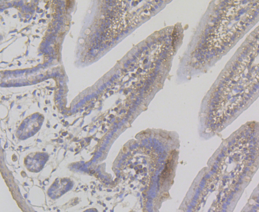
Fig8: Immunohistochemical analysis of paraffin-embedded mouse colon tissue using anti-TRPA1 antibody. The section was pre-treated using heat mediated antigen retrieval with Tris-EDTA buffer (pH 8.0-8.4) for 20 minutes.The tissues were blocked in 5% BSA for 30 minutes at room temperature, washed with ddH2O and PBS, and then probed with the antibody (ER1803-91) at 1/50 dilution, for 30 minutes at room temperature and detected using an HRP conjugated compact polymer system. DAB was used as the chrogen. Counter stained with hematoxylin and mounted with DPX.Application
Fig8: Immunohistochemical analysis of paraffin-embedded mouse colon tissue using anti-TRPA1 antibody. The section was pre-treated using heat mediated antigen retrieval with Tris-EDTA buffer (pH 8.0-8.4) for 20 minutes.The tissues were blocked in 5% BSA for 30 minutes at room temperature, washed with ddH2O and PBS, and then probed with the antibody (ER1803-91) at 1/50 dilution, for 30 minutes at room temperature and detected using an HRP conjugated compact polymer system. DAB was used as the chrogen. Counter stained with hematoxylin and mounted with DPX.Application
Fig8: Immunohistochemical analysis of paraffin-embedded mouse colon tissue using anti-TRPA1 antibody. The section was pre-treated using heat mediated antigen retrieval with Tris-EDTA buffer (pH 8.0-8.4) for 20 minutes.The tissues were blocked in 5% BSA for 30 minutes at room temperature, washed with ddH2O and PBS, and then probed with the antibody (ER1803-91) at 1/50 dilution, for 30 minutes at room temperature and detected using an HRP conjugated compact polymer system. DAB was used as the chrogen. Counter stained with hematoxylin and mounted with DPX.Application
Fig8: Immunohistochemical analysis of paraffin-embedded mouse colon tissue using anti-TRPA1 antibody. The section was pre-treated using heat mediated antigen retrieval with Tris-EDTA buffer (pH 8.0-8.4) for 20 minutes.The tissues were blocked in 5% BSA for 30 minutes at room temperature, washed with ddH2O and PBS, and then probed with the antibody (ER1803-91) at 1/50 dilution, for 30 minutes at room temperature and detected using an HRP conjugated compact polymer system. DAB was used as the chrogen. Counter stained with hematoxylin and mounted with DPX.Application
Fig8: Immunohistochemical analysis of paraffin-embedded mouse colon tissue using anti-TRPA1 antibody. The section was pre-treated using heat mediated antigen retrieval with Tris-EDTA buffer (pH 8.0-8.4) for 20 minutes.The tissues were blocked in 5% BSA for 30 minutes at room temperature, washed with ddH2O and PBS, and then probed with the antibody (ER1803-91) at 1/50 dilution, for 30 minutes at room temperature and detected using an HRP conjugated compact polymer system. DAB was used as the chrogen. Counter stained with hematoxylin and mounted with DPX.Application
Fig8: Immunohistochemical analysis of paraffin-embedded mouse colon tissue using anti-TRPA1 antibody. The section was pre-treated using heat mediated antigen retrieval with Tris-EDTA buffer (pH 8.0-8.4) for 20 minutes.The tissues were blocked in 5% BSA for 30 minutes at room temperature, washed with ddH2O and PBS, and then probed with the antibody (ER1803-91) at 1/50 dilution, for 30 minutes at room temperature and detected using an HRP conjugated compact polymer system. DAB was used as the chrogen. Counter stained with hematoxylin and mounted with DPX.Application
Fig8: Immunohistochemical analysis of paraffin-embedded mouse colon tissue using anti-TRPA1 antibody. The section was pre-treated using heat mediated antigen retrieval with Tris-EDTA buffer (pH 8.0-8.4) for 20 minutes.The tissues were blocked in 5% BSA for 30 minutes at room temperature, washed with ddH2O and PBS, and then probed with the antibody (ER1803-91) at 1/50 dilution, for 30 minutes at room temperature and detected using an HRP conjugated compact polymer system. DAB was used as the chrogen. Counter stained with hematoxylin and mounted with DPX.Application
Fig8: Immunohistochemical analysis of paraffin-embedded mouse colon tissue using anti-TRPA1 antibody. The section was pre-treated using heat mediated antigen retrieval with Tris-EDTA buffer (pH 8.0-8.4) for 20 minutes.The tissues were blocked in 5% BSA for 30 minutes at room temperature, washed with ddH2O and PBS, and then probed with the antibody (ER1803-91) at 1/50 dilution, for 30 minutes at room temperature and detected using an HRP conjugated compact polymer system. DAB was used as the chrogen. Counter stained with hematoxylin and mounted with DPX.IHC
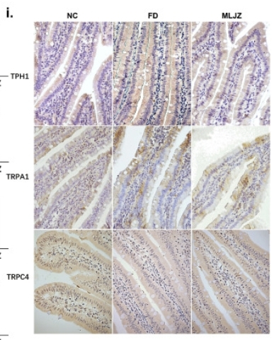
i) IHC staining for TPH1, TRPA1, TRPC4 in rats duodenumIF
b Immunofluorescence staining of TRPA1 in OLN-93 oligodendrocyte. DAPI (blue), MBP-AF594 (red), and TRPA1-AF488 (green) are shown.| Product Name | TRPA1 |
|---|---|
| Antibody Type | Primary Antibodies |
| Product description | Receptor-activated non-selective cation channel involved in detection of pain and possibly also in cold perception and inner ear function. Has a central role in the pain response to endogenous inflammatory mediators and to a diverse array of volatile irritants, such as mustard oil, cinnamaldehyde, garlic and acrolein, an irritant from tears gas and vehicule exhaust fumes. Is also activated by menthol (in vitro). Acts also as a ionotropic cannabinoid receptor by being activated by delta9-tetrahydrocannabinol (THC), the psychoactive component of marijuana. May be a component for the mechanosensitive transduction channel of hair cells in inner ear, thereby participating in the perception of sounds. Probably operated by a phosphatidylinositol second messenger system. |
| Immunogen | Recombinant protein within Human TRPA1 aa 500-720. |
| Clonality | Polyclonal |
|---|---|
| Isotype | IgG |
| Host Species | Rabbit |
| WB:1:500-1:1,000: ICC:1:50-1:200: IHC:1:50-1:200: |
|
| Concentration | 1mg/ml |
| Alternative Names | ANKTM 1 antibody ANKTM1 antibody Ankyrin-like with transmembrane domains protein 1 antibody Transformation sensitive protein p120 antibody Transformation-sensitive protein p120 antibody Transient receptor potential cation channel subfamily A member 1 antibody TRPA 1 antibody Trpa1 antibody TRPA1_HUMAN antibody |
|---|---|
| Molecular Weight(MW) | Predicted band size: 127 kDa |
| Cellular Localization | Cell membrane. |
Application
Fig8: Immunohistochemical analysis of paraffin-embedded mouse colon tissue using anti-TRPA1 antibody. The section was pre-treated using heat mediated antigen retrieval with Tris-EDTA buffer (pH 8.0-8.4) for 20 minutes.The tissues were blocked in 5% BSA for 30 minutes at room temperature, washed with ddH2O and PBS, and then probed with the antibody (ER1803-91) at 1/50 dilution, for 30 minutes at room temperature and detected using an HRP conjugated compact polymer system. DAB was used as the chrogen. Counter stained with hematoxylin and mounted with DPX.
Application
Fig8: Immunohistochemical analysis of paraffin-embedded mouse colon tissue using anti-TRPA1 antibody. The section was pre-treated using heat mediated antigen retrieval with Tris-EDTA buffer (pH 8.0-8.4) for 20 minutes.The tissues were blocked in 5% BSA for 30 minutes at room temperature, washed with ddH2O and PBS, and then probed with the antibody (ER1803-91) at 1/50 dilution, for 30 minutes at room temperature and detected using an HRP conjugated compact polymer system. DAB was used as the chrogen. Counter stained with hematoxylin and mounted with DPX.
Application
Fig8: Immunohistochemical analysis of paraffin-embedded mouse colon tissue using anti-TRPA1 antibody. The section was pre-treated using heat mediated antigen retrieval with Tris-EDTA buffer (pH 8.0-8.4) for 20 minutes.The tissues were blocked in 5% BSA for 30 minutes at room temperature, washed with ddH2O and PBS, and then probed with the antibody (ER1803-91) at 1/50 dilution, for 30 minutes at room temperature and detected using an HRP conjugated compact polymer system. DAB was used as the chrogen. Counter stained with hematoxylin and mounted with DPX.
Application
Fig8: Immunohistochemical analysis of paraffin-embedded mouse colon tissue using anti-TRPA1 antibody. The section was pre-treated using heat mediated antigen retrieval with Tris-EDTA buffer (pH 8.0-8.4) for 20 minutes.The tissues were blocked in 5% BSA for 30 minutes at room temperature, washed with ddH2O and PBS, and then probed with the antibody (ER1803-91) at 1/50 dilution, for 30 minutes at room temperature and detected using an HRP conjugated compact polymer system. DAB was used as the chrogen. Counter stained with hematoxylin and mounted with DPX.
Application
Fig8: Immunohistochemical analysis of paraffin-embedded mouse colon tissue using anti-TRPA1 antibody. The section was pre-treated using heat mediated antigen retrieval with Tris-EDTA buffer (pH 8.0-8.4) for 20 minutes.The tissues were blocked in 5% BSA for 30 minutes at room temperature, washed with ddH2O and PBS, and then probed with the antibody (ER1803-91) at 1/50 dilution, for 30 minutes at room temperature and detected using an HRP conjugated compact polymer system. DAB was used as the chrogen. Counter stained with hematoxylin and mounted with DPX.
Application
Fig8: Immunohistochemical analysis of paraffin-embedded mouse colon tissue using anti-TRPA1 antibody. The section was pre-treated using heat mediated antigen retrieval with Tris-EDTA buffer (pH 8.0-8.4) for 20 minutes.The tissues were blocked in 5% BSA for 30 minutes at room temperature, washed with ddH2O and PBS, and then probed with the antibody (ER1803-91) at 1/50 dilution, for 30 minutes at room temperature and detected using an HRP conjugated compact polymer system. DAB was used as the chrogen. Counter stained with hematoxylin and mounted with DPX.
Application
Fig8: Immunohistochemical analysis of paraffin-embedded mouse colon tissue using anti-TRPA1 antibody. The section was pre-treated using heat mediated antigen retrieval with Tris-EDTA buffer (pH 8.0-8.4) for 20 minutes.The tissues were blocked in 5% BSA for 30 minutes at room temperature, washed with ddH2O and PBS, and then probed with the antibody (ER1803-91) at 1/50 dilution, for 30 minutes at room temperature and detected using an HRP conjugated compact polymer system. DAB was used as the chrogen. Counter stained with hematoxylin and mounted with DPX.
Application
Fig8: Immunohistochemical analysis of paraffin-embedded mouse colon tissue using anti-TRPA1 antibody. The section was pre-treated using heat mediated antigen retrieval with Tris-EDTA buffer (pH 8.0-8.4) for 20 minutes.The tissues were blocked in 5% BSA for 30 minutes at room temperature, washed with ddH2O and PBS, and then probed with the antibody (ER1803-91) at 1/50 dilution, for 30 minutes at room temperature and detected using an HRP conjugated compact polymer system. DAB was used as the chrogen. Counter stained with hematoxylin and mounted with DPX.
IHC
i) IHC staining for TPH1, TRPA1, TRPC4 in rats duodenum
IF
b Immunofluorescence staining of TRPA1 in OLN-93 oligodendrocyte. DAPI (blue), MBP-AF594 (red), and TRPA1-AF488 (green) are shown.| Positive Control | L929, MG-63, NIH-3T3, rat brain tissue, human skin tissue, human esophagus tissue, mouse colon tissue. |
|---|---|
| Application Notes | WB:1:500-1:1,000: ICC:1:50-1:200: IHC:1:50-1:200: |
| Form | Liquid |
|---|---|
| Storage Instructions | Store at +4℃ after thawing. Aliquot store at -20℃. Avoid repeated freeze / thaw cycles. |
| Storage Buffer | 1*TBS (pH7.4), 1%BSA, 50%Glycerol. Preservative: 0.05% Sodium Azide. |


